Copyright
©The Author(s) 2019.
World J Clin Cases. Aug 6, 2019; 7(15): 2094-2102
Published online Aug 6, 2019. doi: 10.12998/wjcc.v7.i15.2094
Published online Aug 6, 2019. doi: 10.12998/wjcc.v7.i15.2094
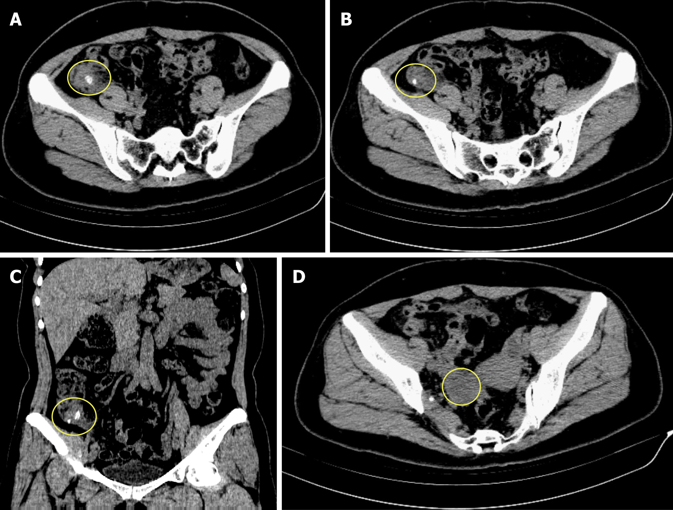
Figure 1 Computed tomography scan of the patient’s abdomen.
A and B: Transverse sections showing swelling of the appendix with surrounding exudation, and there is a fecalith at the root of the appendix, respectively; C: Coronal section showing swelling of the appendix with exudation and two separate fecaliths at its root; D: Transverse section showing a low density mass located in the right ovary.
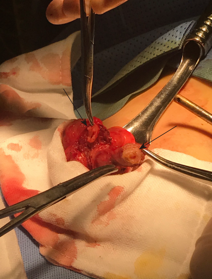
Figure 2 Intraoperative photograph of the patient.
The intraoperative photograph shows that there are two bases of the appendix at the end of the cecum, and both of these bases are in contact with the cecum (black arrow).
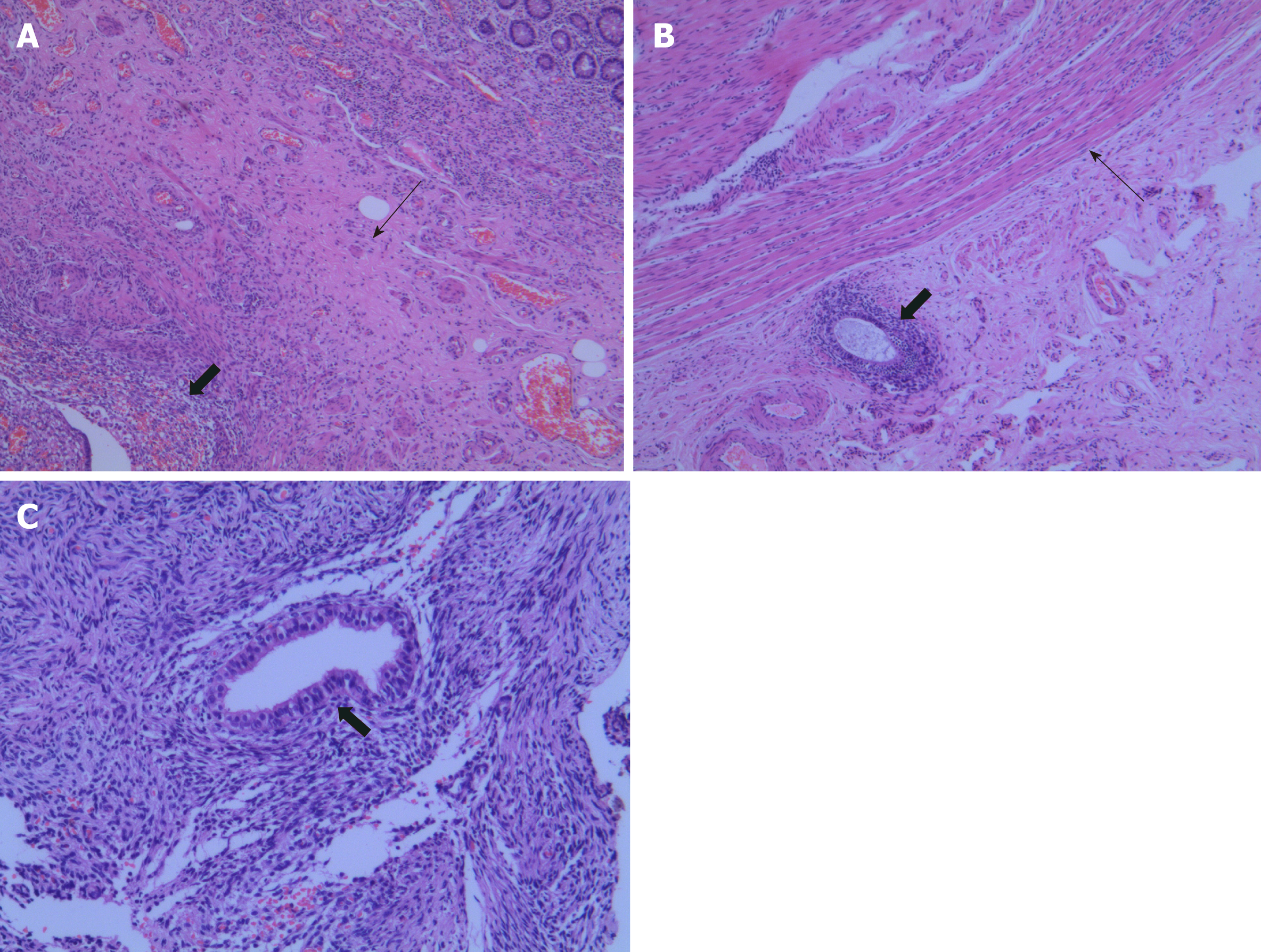
Figure 3 Histopathological appearances of the endometriosis in appendix, small intestine, and ovary.
A: Hematoxylin and eosin (HE) staining shows that ectopic endometrial-type glands and stroma (thick arrows) were detected in the muscular layers (thin arrows) of the appendix (×40); B: Ectopic endometrial-type glands and stroma (thick arrows) were observed in the subserosa of the small intestine. The muscular layers are indicated by thin arrows (HE, ×40); C: Higher magnification of endometriosis (thick arrows) in the ovary (HE, ×100).
- Citation: Zhu MY, Fei FM, Chen J, Zhou ZC, Wu B, Shen YY. Endometriosis of the duplex appendix: A case report and review of the literature. World J Clin Cases 2019; 7(15): 2094-2102
- URL: https://www.wjgnet.com/2307-8960/full/v7/i15/2094.htm
- DOI: https://dx.doi.org/10.12998/wjcc.v7.i15.2094


