Copyright
©The Author(s) 2019.
World J Clin Cases. Jul 26, 2019; 7(14): 1899-1907
Published online Jul 26, 2019. doi: 10.12998/wjcc.v7.i14.1899
Published online Jul 26, 2019. doi: 10.12998/wjcc.v7.i14.1899
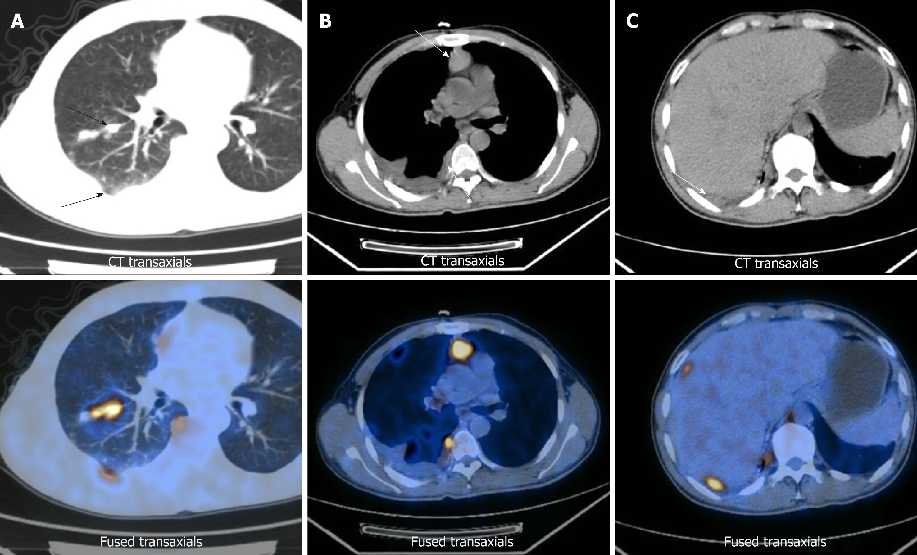
Figure 1 Images from positron emission tomography/computed tomography preformed on April 12, 2013.
A: The primary tumor and pleural metastasis; B: Mediastinal metastasis; C: Pleural metastasis.
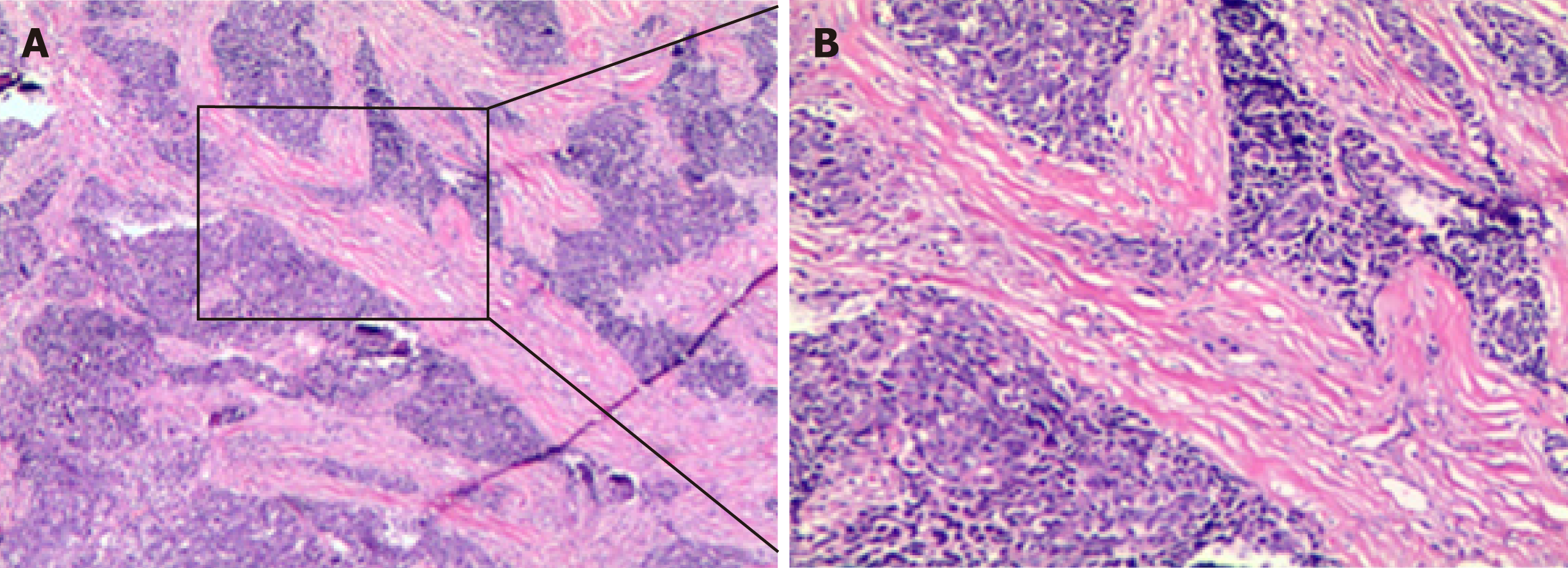
Figure 2 Pathological findings on April 23, 2013.
The biopsied sample (hematoxylin-eosin staining) showed lowly-differentiated squamous cell carcinoma of the chest wall (derived from the right lung). Scale bar represents 100 μm.
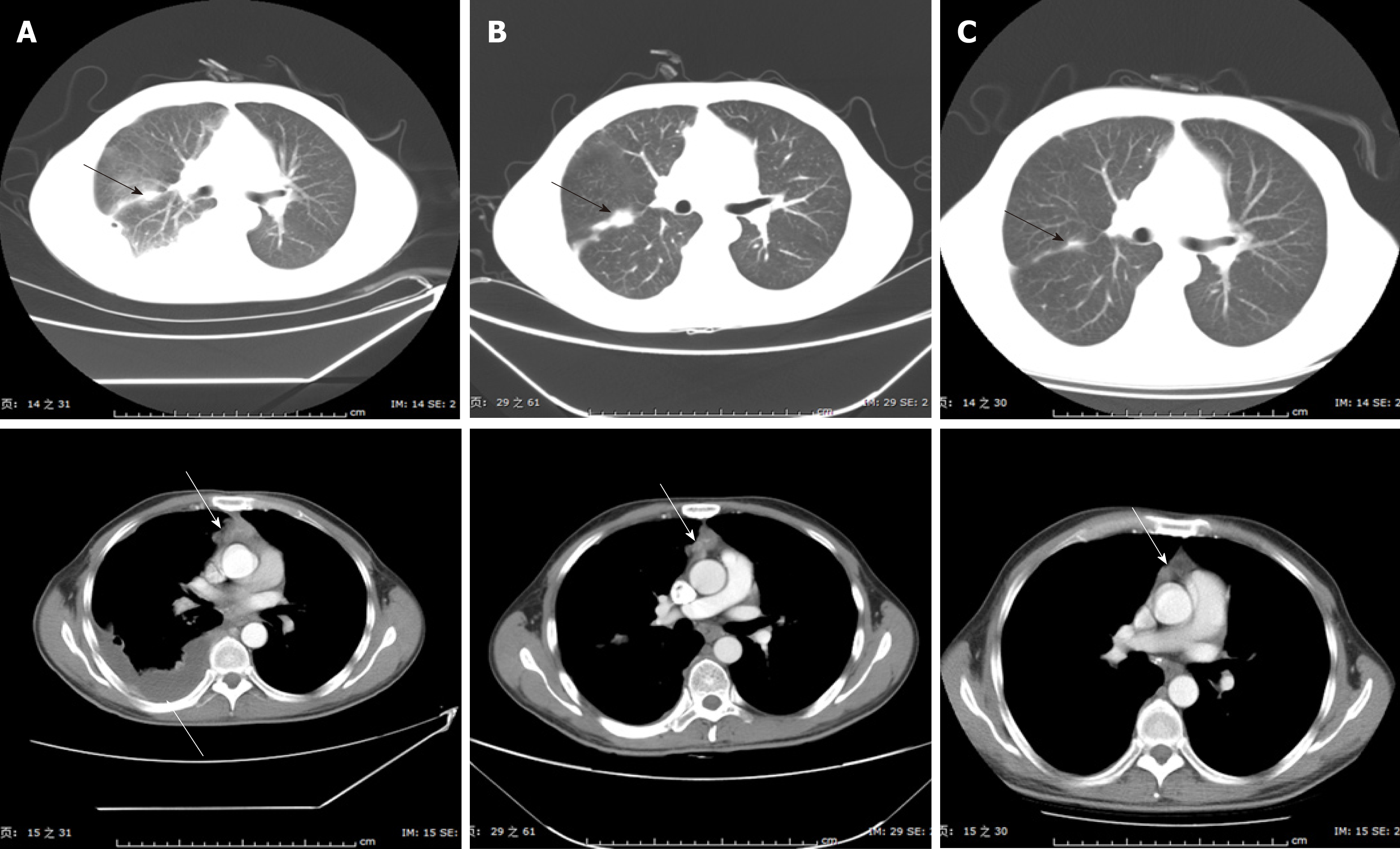
Figure 3 Images from computed tomography showing the tumor in the right lung and mediastinum in response to therapy.
A: Before therapy, imaging performed on May 2, 2013; B: After two cycles of DP therapy, imaging performed on June 7, 2013 and evaluation classified the status as a progressive disease; C: After six cycles of TP therapy, imaging showed that the tumor had shrank significantly. DP: 75 mg/m2 docetaxel on day 1 and 75 mg/m2 cisplatin on day 1; TP: 100 mg/m2 nab-paclitaxel (nab-PC) on days 1, 8 ,and 15, and 75 mg/m2 cisplatin on day 1.
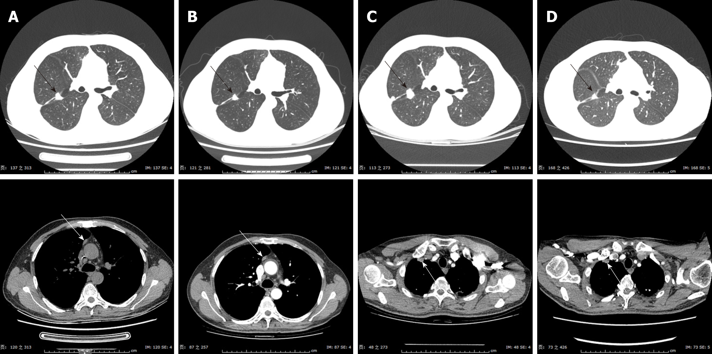
Figure 4 Images from computed tomography showing the tumor response up to the fourth-line therapy of NVB + Endo.
A: Imaging performed on May 17, 2016, after seven cycles of nab-PC therapy; B: On December 26, 2016, the disease was classified as partial remission; C: On April 17, 2017, the disease was classified as progressive; D: On April 27, 2017, the tumor was found to have a response to radiation therapy in the right lung (DT = 50 Gy/5F). Nab-PC: Nab-paclitaxel; NVB + Endo: 25 mg/m2 vinorelbine on days 1 and 8, and 30 mg endostar on days 1-7.
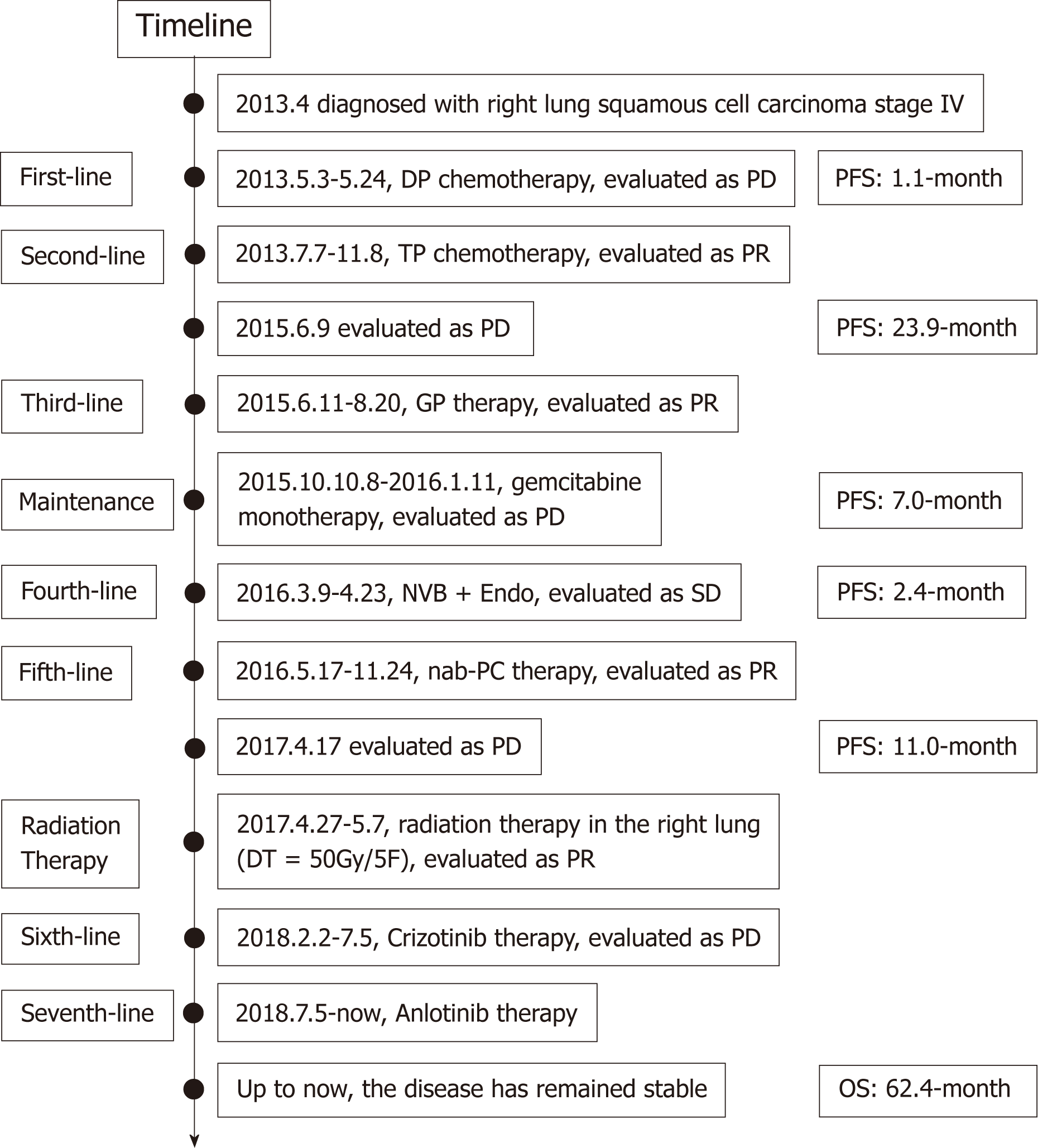
Figure 5 Timeline of our case’s treatment with multiline therapy for advanced squamous cell carcinoma.
- Citation: Yang X, Peng P, Zhang L. Multiline treatment of advanced squamous cell carcinoma of the lung: A case report and review of the literature. World J Clin Cases 2019; 7(14): 1899-1907
- URL: https://www.wjgnet.com/2307-8960/full/v7/i14/1899.htm
- DOI: https://dx.doi.org/10.12998/wjcc.v7.i14.1899


