Copyright
©The Author(s) 2019.
World J Clin Cases. Jul 6, 2019; 7(13): 1671-1676
Published online Jul 6, 2019. doi: 10.12998/wjcc.v7.i13.1671
Published online Jul 6, 2019. doi: 10.12998/wjcc.v7.i13.1671
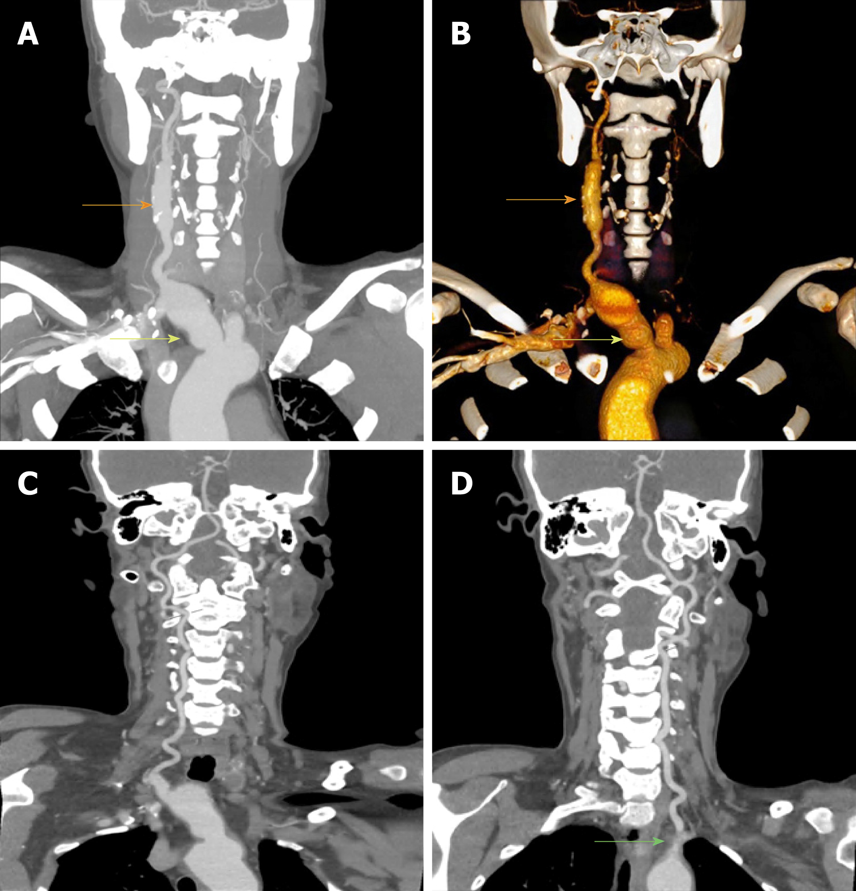
Figure 1 Preoperative computed tomography angiography of the patient.
A and B: Innominate (yellow arrow) and right common carotid (orange arrow) arterial aneurysms and no display of the left carotid and subclavian arteries; C: Patency of the right vertebral artery; D: Occlusion of the initial part of the left vertebral artery (green arrow).
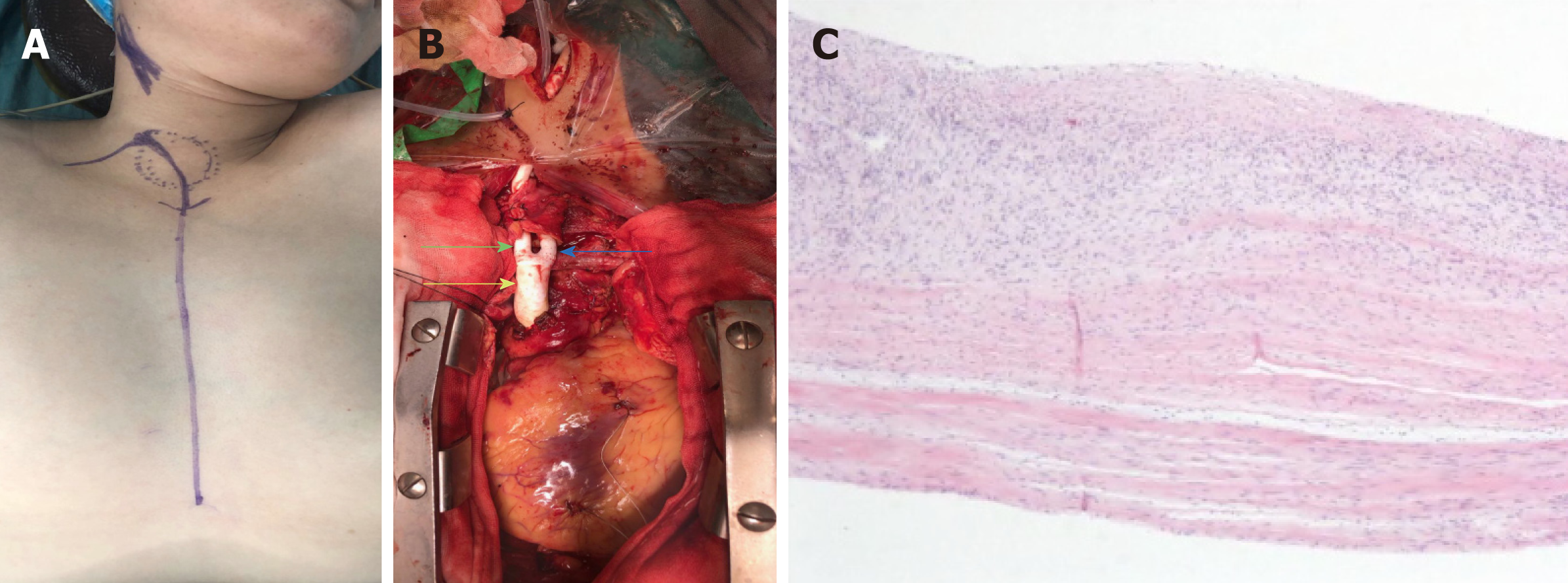
Figure 2 Macroscopic photograph of surgery and hematoxylin-eosin staining of vascular tissue.
A: The main thoracic incision; B: The replaced artificial vessel (green arrow: subclavian end; blue arrow: carotid end; yellow arrow: aortic end); C: Hematoxylin-eosin staining of vascular tissue excised.
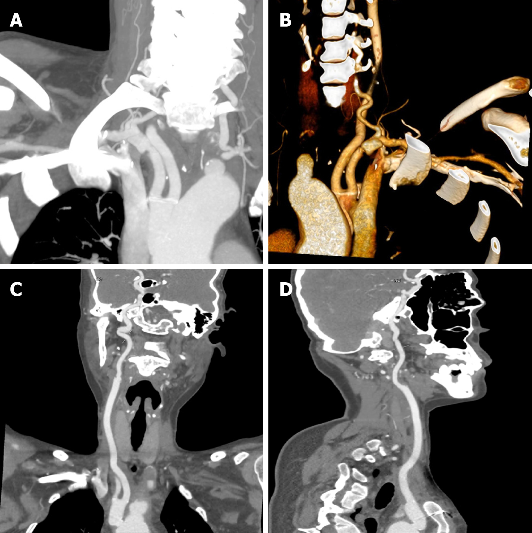
Figure 3 Computed tomography angiography images obtained 1 mo after surgery demonstrating patency of the artificial vessel with no dilatation.
- Citation: Wang WD, Sun R, Zhou MX, Liu XR, Zheng YH, Chen YX. A complicated case of innominate and right common arterial aneurysms due to Takayasu’s arteritis. World J Clin Cases 2019; 7(13): 1671-1676
- URL: https://www.wjgnet.com/2307-8960/full/v7/i13/1671.htm
- DOI: https://dx.doi.org/10.12998/wjcc.v7.i13.1671


