Copyright
©The Author(s) 2019.
World J Clin Cases. Jan 6, 2019; 7(1): 95-101
Published online Jan 6, 2019. doi: 10.12998/wjcc.v7.i1.95
Published online Jan 6, 2019. doi: 10.12998/wjcc.v7.i1.95
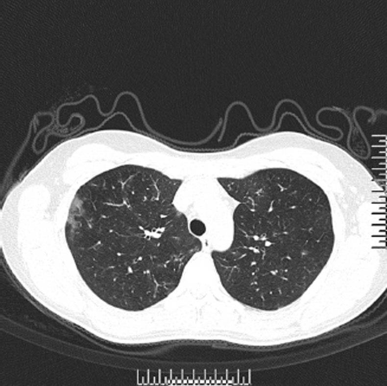
Figure 1 High resolution axial computed tomography image showing bilateral bronchial wall thickening accompanied with patchy spots scattered throughout bilateral lungs.
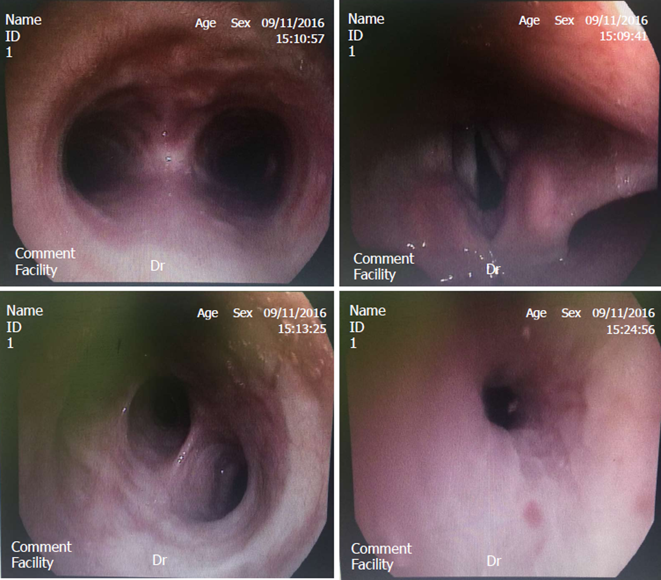
Figure 2 Bronchofiberscopic images showing that the bronchial mucosa had hyperemia, oedema and congestion, especially in the right superior lobe.
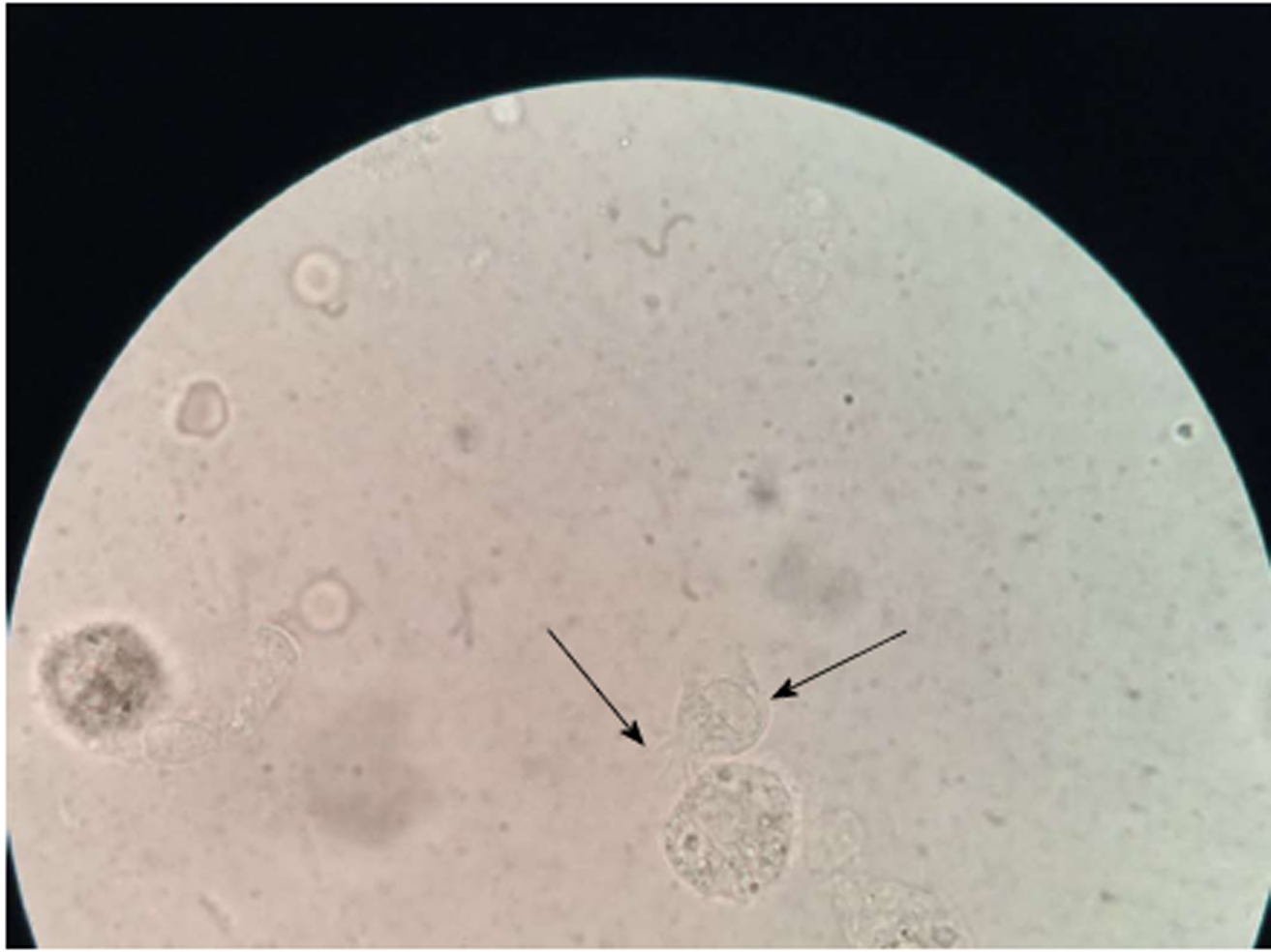
Figure 3 Living cells with cilia in bronchoalveolar lavage fluid.
Left arrow: The cilia on the top of the cell oscillate rapidly to drive cell migration; Right arrow: The body of the cell.
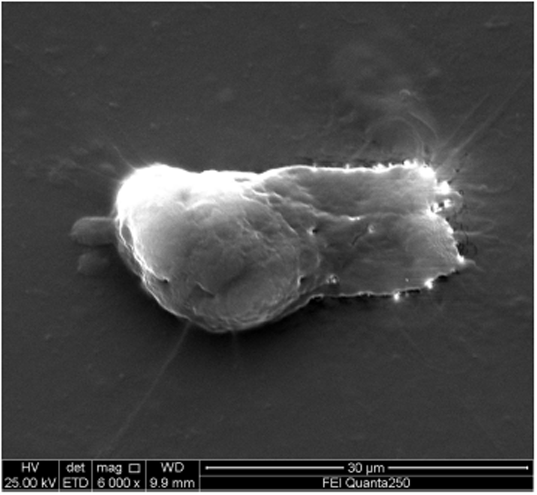
Figure 4 Morphological characteristics under a scanning electron microscope.
The nucleus of ciliary active cells is far away from the ciliate tip of the cell, and is located at the bottom of the cell.
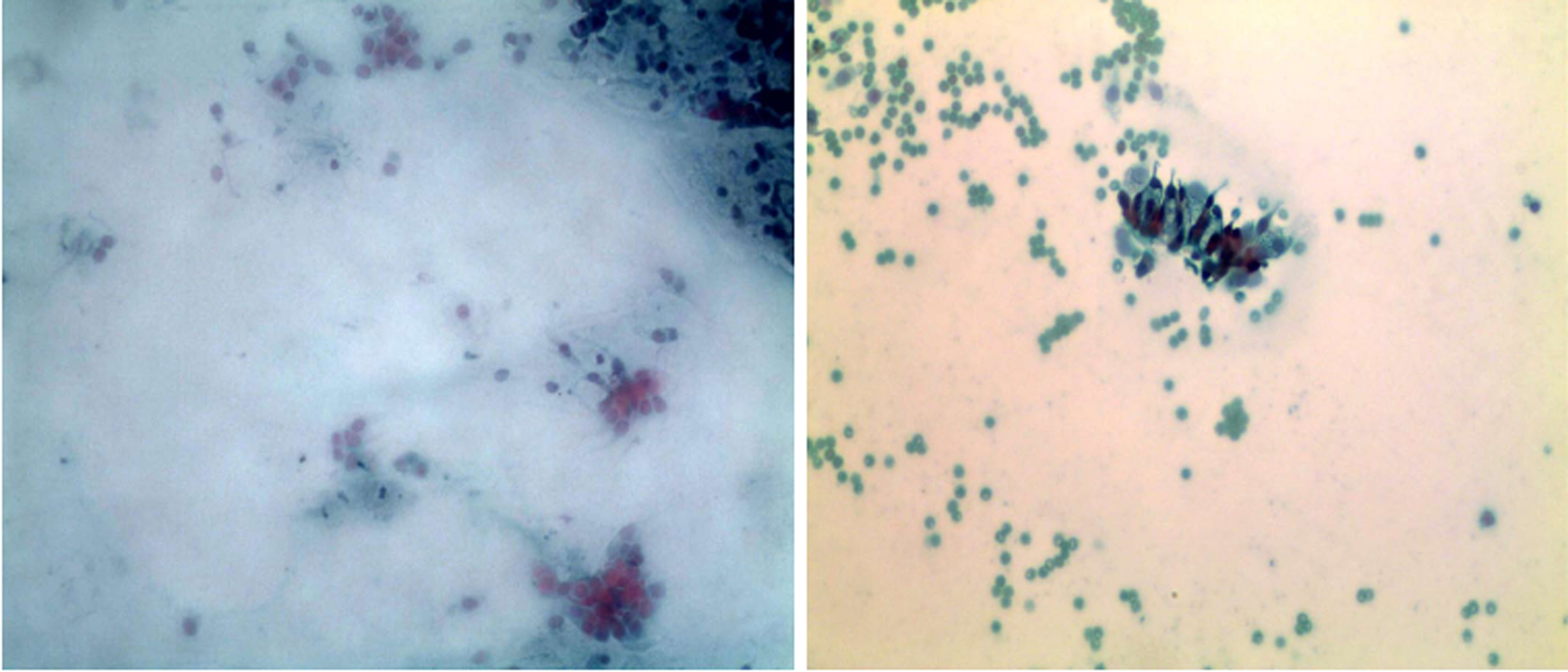
Figure 5 Brush smears stained with Pap stain.
Numerous scattered or gathered respiratory ciliated cells are visible.
- Citation: Meng SS, Dai ZF, Wang HC, Li YX, Wei DD, Yang RL, Lin XH. Authenticity of pulmonary Lophomonas blattarum infection: A case report. World J Clin Cases 2019; 7(1): 95-101
- URL: https://www.wjgnet.com/2307-8960/full/v7/i1/95.htm
- DOI: https://dx.doi.org/10.12998/wjcc.v7.i1.95


