Copyright
©The Author(s) 2019.
World J Clin Cases. Jan 6, 2019; 7(1): 58-68
Published online Jan 6, 2019. doi: 10.12998/wjcc.v7.i1.58
Published online Jan 6, 2019. doi: 10.12998/wjcc.v7.i1.58
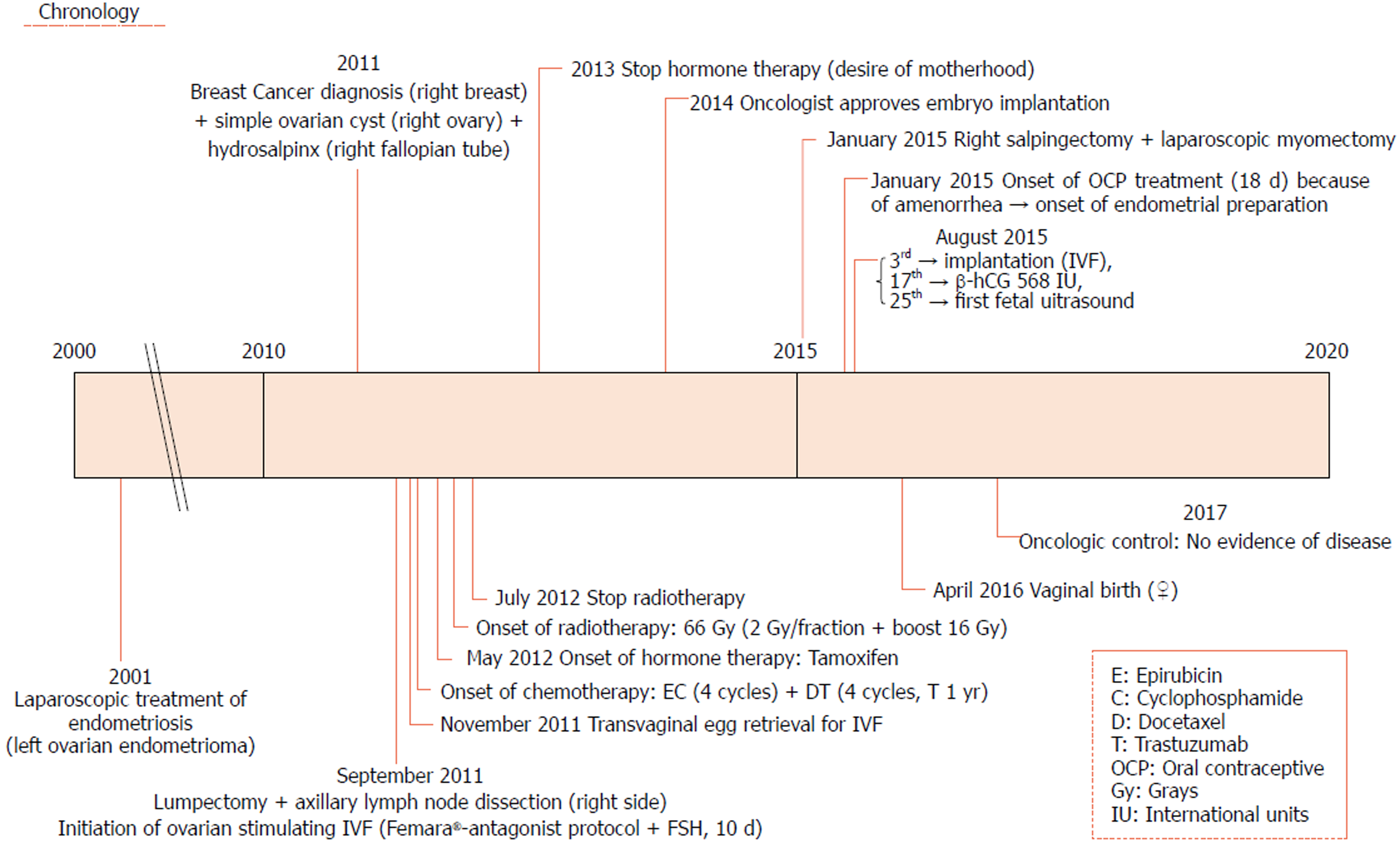
Figure 1 Chronological axis of the patient’s medical history.
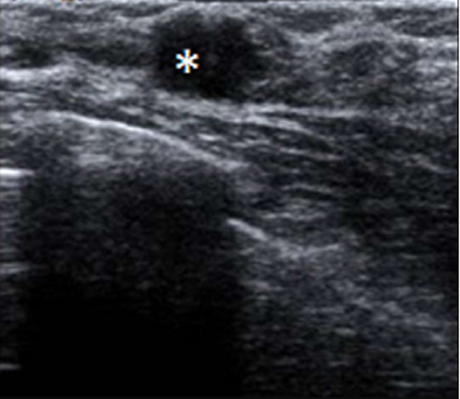
Figure 2 Mammary ultrasound image.
Asterisk showing solid tumour, badly defined, irregular and blurry borders with light posterior shadow. Signs of malignancy on ultrasound.
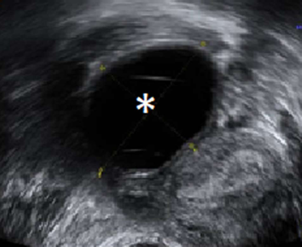
Figure 3 Transvaginal gynecological ultrasound.
Asterisk shows ovarian cystic tumour containing clear liquid, well-defined borders, and smooth internal walls, suggesting serous ovarian cyst.
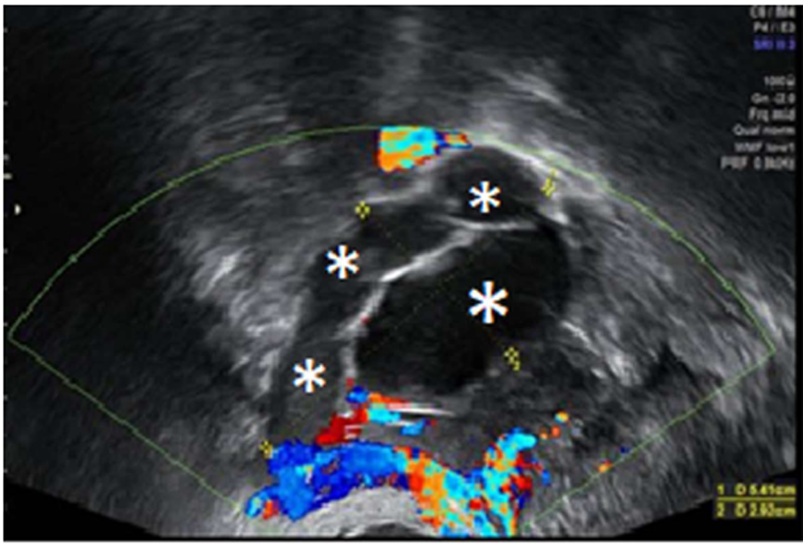
Figure 4 Transvaginal gynecological ultrasound.
Asterisks show complex paraovarian tumor, multilobed, containing liquid and semiliquid, well defined, and with internal walls, suggesting hydrosalpinx. Doppler map of low vascularization and Doppler fluxometry with normal resistances.
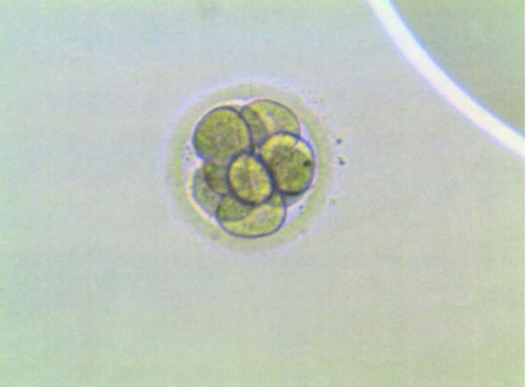
Figure 5 Type A embryo transferred to the patient.
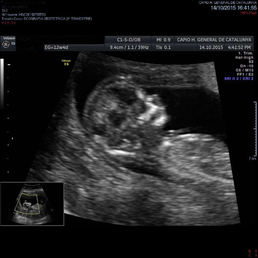
Figure 6 Abdominal obstetric ultrasound.
Dorso-posterior feed with first fetal position of a second trimester fetus.
- Citation: Garrido-Marín M, Argacha PM, Fernández L, Molfino F, Martínez-Soler F, Tortosa A, Gimenez-Bonafé P. Full-term pregnancy in breast cancer survivor with fertility preservation: A case report and review of literature. World J Clin Cases 2019; 7(1): 58-68
- URL: https://www.wjgnet.com/2307-8960/full/v7/i1/58.htm
- DOI: https://dx.doi.org/10.12998/wjcc.v7.i1.58


