Copyright
©The Author(s) 2018.
World J Clin Cases. Sep 6, 2018; 6(9): 274-278
Published online Sep 6, 2018. doi: 10.12998/wjcc.v6.i9.274
Published online Sep 6, 2018. doi: 10.12998/wjcc.v6.i9.274
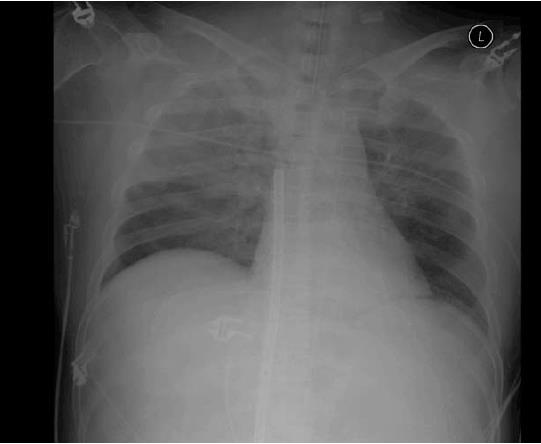
Figure 1 Chest X-ray in the critical care unit.
Right upper lung field infiltration is visible. There is no central venous catheter.
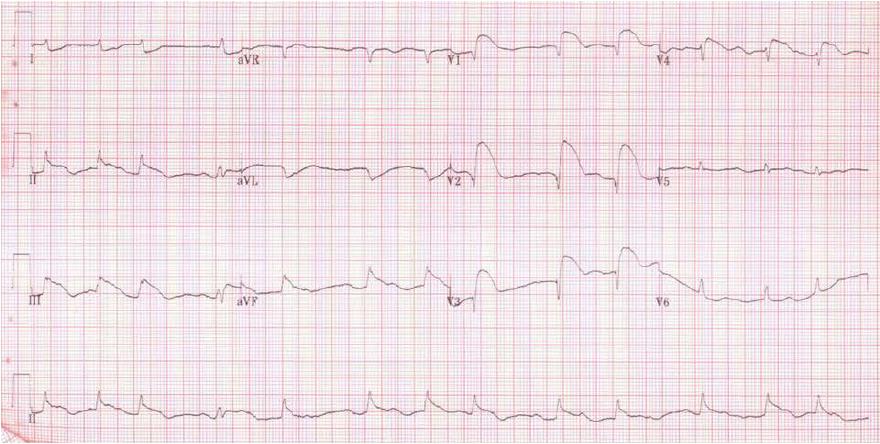
Figure 2 Electrocardiogram after the development of sudden hypotension and loss of consciousness.
The electrocardiogram shows inferior wall infarction.
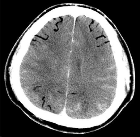
Figure 3 Brain computed tomography image after the development of sudden hypotension and loss of consciousness.
A massive cerebral air embolism is observed.
- Citation: Ryu SM, Park SM. Unexpected complication during extracorporeal membrane oxygenation support: Ventilator associated systemic air embolism. World J Clin Cases 2018; 6(9): 274-278
- URL: https://www.wjgnet.com/2307-8960/full/v6/i9/274.htm
- DOI: https://dx.doi.org/10.12998/wjcc.v6.i9.274


