Copyright
©The Author(s) 2018.
World J Clin Cases. Sep 6, 2018; 6(9): 249-258
Published online Sep 6, 2018. doi: 10.12998/wjcc.v6.i9.249
Published online Sep 6, 2018. doi: 10.12998/wjcc.v6.i9.249
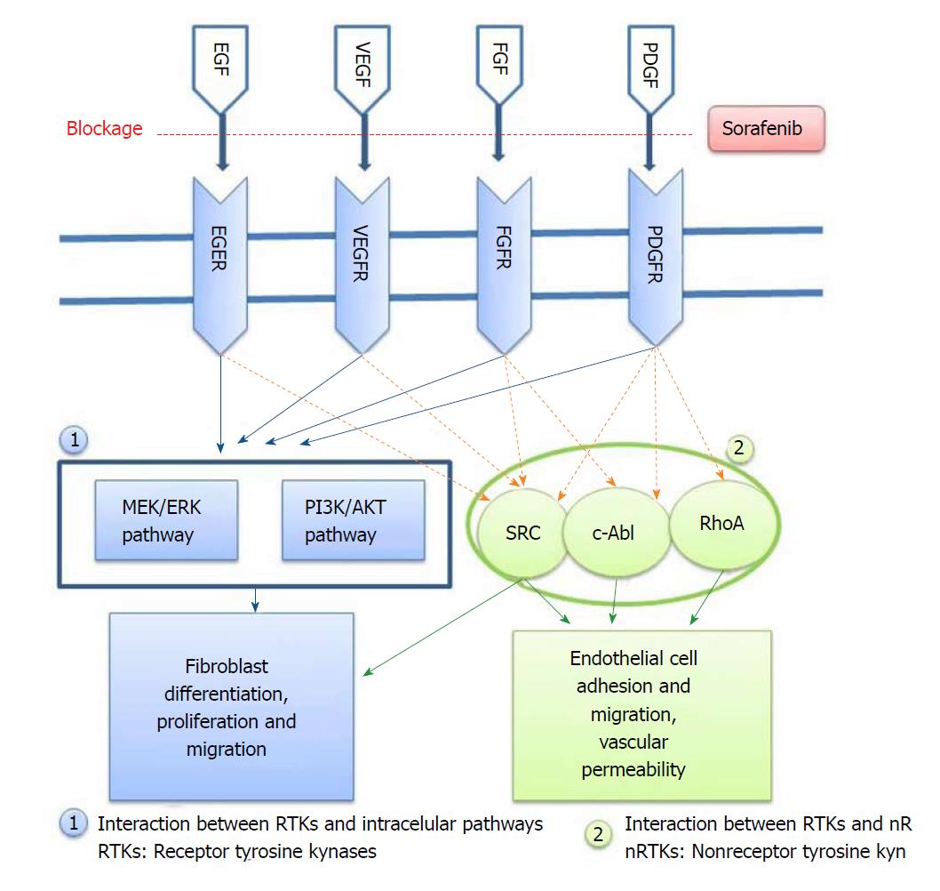
Figure 1 Fibrosis development mechanism and the stage at which sorafenib prevents fibrosis is indicated.
VEGF: Vascular endothelial growth factor.
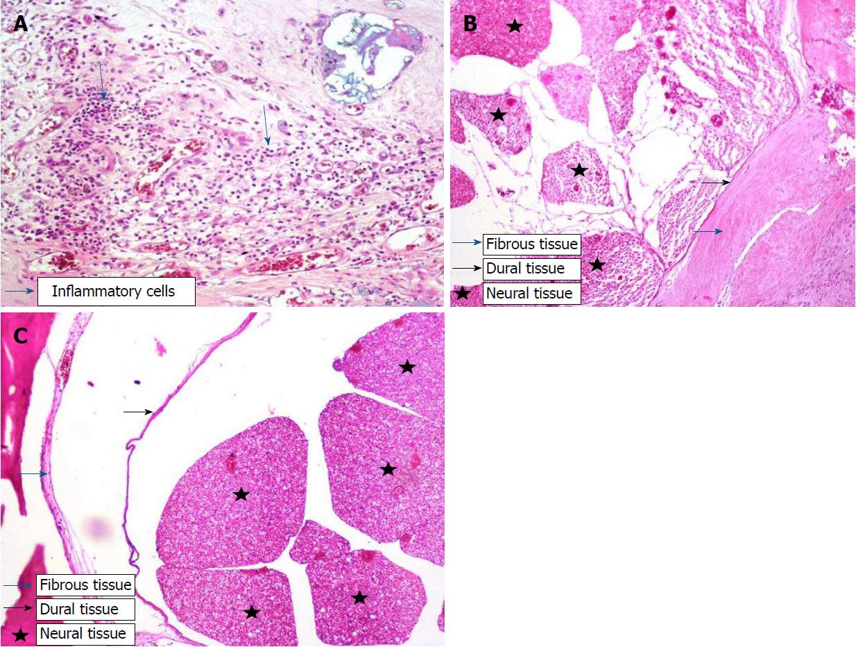
Figure 2 Histopathological tissue section stained by HE.
A: Grade-3 fibrosis and inflammatory cells in Group I; B: Grade-3 fibrosis and fibrous tissue in Group I; C: Grade 1 fibrosis in Group II. Star: Neural tissues; black arrow: Dura; blue arrow: Fibrosis tissue.
Figure 3 Immunohistochemical tissue section stained with anti-CD105 antibody.
A: Grade-3 fibrosis in Group I; B: Grade-1 fibrosis in Group II. blue arrow: Fibrosis.
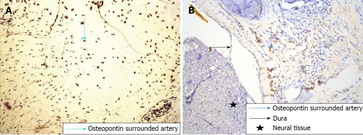
Figure 4 Immunohistochemical tissue section stained with anti-osteopontin antibody.
A: Grade-3 fibrosis in Group I; B: Grade-1 fibrosis in Group II. Star: Neural tissues; black arrow: Dura; blue arrow: Fibrosis tissue.
- Citation: Tanriverdi O, Erdogan U, Tanik C, Yilmaz I, Gunaldi O, Adilay HU, Arslanhan A, Eseoglu M. Impact of sorafenib on epidural fibrosis: An immunohistochemical study. World J Clin Cases 2018; 6(9): 249-258
- URL: https://www.wjgnet.com/2307-8960/full/v6/i9/249.htm
- DOI: https://dx.doi.org/10.12998/wjcc.v6.i9.249


