Copyright
©The Author(s) 2018.
World J Clin Cases. Jul 16, 2018; 6(7): 150-155
Published online Jul 16, 2018. doi: 10.12998/wjcc.v6.i7.150
Published online Jul 16, 2018. doi: 10.12998/wjcc.v6.i7.150
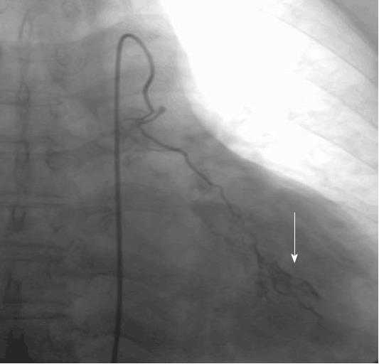
Figure 1 Selective arterial angiography showing notable contrast agent diffusion in the left lower lung, indicating blood supply to the tumor (white arrow).
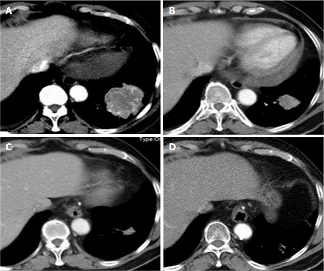
Figure 2 Transverse computed tomography scan findings of patients before and during treatment.
A: Pre-treatment transverse computed tomography (CT) scan showing the tumor mass in the basal segment of the left lower lobe. The greatest dimension of the tumor measured 5.7 cm; B: One month after treatment, transverse CT scan showed significant reduction in tumor size. The greatest dimension measured 3.5 cm; C: Seven months after treatment, CT scan showed further significant reduction in tumor size. Greatest dimension was reduced to 1.8 cm; D: CT scan 45 mo after treatment showed that tumor almost disappeared (only fiber scar tissue was found by puncture biopsy).
- Citation: Yang NN, Xiong F, He Q, Guan YS. Achievable complete remission of advanced non-small-cell lung cancer: Case report and review of the literature. World J Clin Cases 2018; 6(7): 150-155
- URL: https://www.wjgnet.com/2307-8960/full/v6/i7/150.htm
- DOI: https://dx.doi.org/10.12998/wjcc.v6.i7.150


