Copyright
©The Author(s) 2018.
World J Clin Cases. Dec 26, 2018; 6(16): 1169-1174
Published online Dec 26, 2018. doi: 10.12998/wjcc.v6.i16.1169
Published online Dec 26, 2018. doi: 10.12998/wjcc.v6.i16.1169
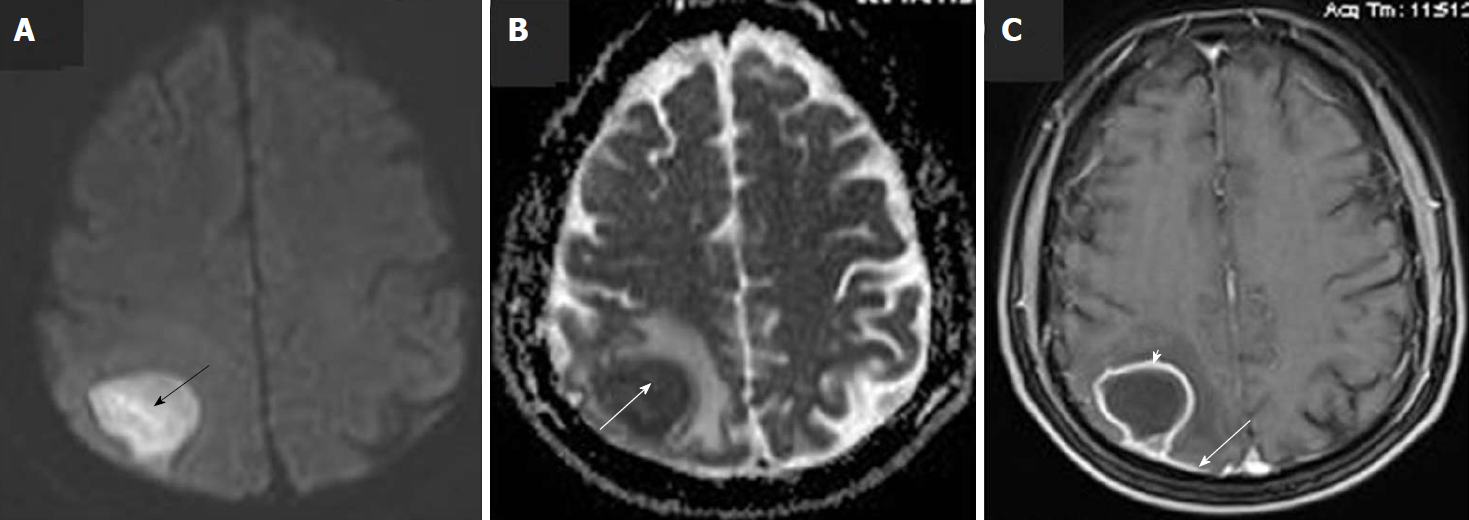
Figure 1 Pre-operative brain magnetic resonance imaging scan with intravenous Gadolinium administration.
A: Axial diffusion-weighted imaging (DWI) image shows high signal of the lesion in right upper parietal lobe (black arrow); B: Axial DWI ADC map reveals low signal at the same area, due to restricted diffusion (white arrow); C: Axial post-Gd image, shows rim enhancement of the lesion (white arrowhead) as well as enhancement of the adjacent dura (white arrow).
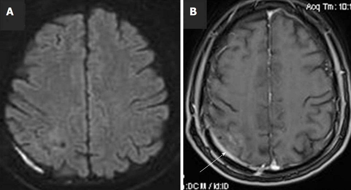
Figure 2 Follow-up brain magnetic resonance imaging scan with intravenous Gadolinium administration, at two months post operation.
A: diffusion-weighted imaging shows complete removal of the lesion, with normal diffusion of the brain parenchyma; B: Axial post-Gd image, shows minimal enhancement of the adjacent dura, due to the previous surgery (white arrow).
- Citation: Tsonis I, Karamani L, Xaplanteri P, Kolonitsiou F, Zampakis P, Gatzounis G, Marangos M, Assimakopoulos SF. Spontaneous cerebral abscess due to Bacillus subtilis in an immunocompetent male patient: A case report and review of literature. World J Clin Cases 2018; 6(16): 1169-1174
- URL: https://www.wjgnet.com/2307-8960/full/v6/i16/1169.htm
- DOI: https://dx.doi.org/10.12998/wjcc.v6.i16.1169


