Copyright
©The Author(s) 2018.
World J Clin Cases. Dec 6, 2018; 6(15): 1018-1023
Published online Dec 6, 2018. doi: 10.12998/wjcc.v6.i15.1018
Published online Dec 6, 2018. doi: 10.12998/wjcc.v6.i15.1018
Figure 1 Initial radiological appearance of the kidney tumor and the thyroid left lobe mass.
A: Preoperative contrast-enhanced computed tomography (CT) scan image of the left kidney tumor. The image shows a 6.0 cm × 5.0 cm exophytic mass in the lower pole of the left kidney (arrowheads). No lymphadenopathy was identified; B: Contrast-enhanced CT scan revealed a 4.0 cm × 2.5 cm solid mass with a smooth surface in the left lobe of the thyroid gland (arrowheads); C: The ultrasound imaging shows a 3.2 cm × 2.8 cm × 3.3 cm hypoechoic mass lesion without calcification in the left lobe of the thyroid.
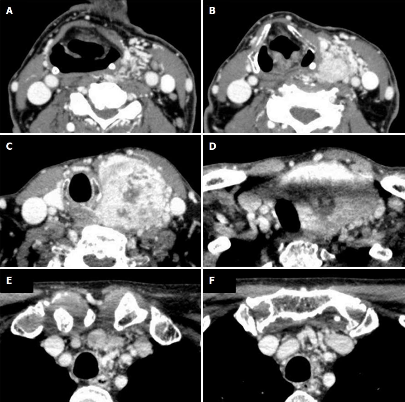
Figure 2 Serial (A-F) contrast-enhanced computed tomography scan images of the thyroid mass 43 mo after the first examination.
The computed tomography scan reveals a heterogeneously contrast-enhanced large tumor surrounded by various vascular-like structures. The trachea was compressed but invasion was not apparent.
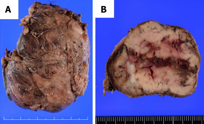
Figure 3 Gross appearance of the resected specimen.
A: The left lobe of the thyroid was markedly enlarged (6.5 cm × 5.0 cm); B: The cut surface of the resected specimen showed a whitish and partially hemorrhagic solid tumor. The tumor did not invade beyond the capsule of the thyroid.
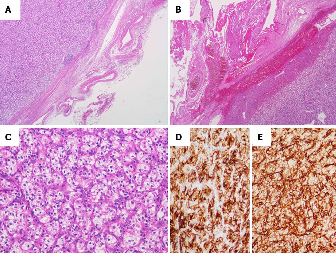
Figure 4 Histological findings of the resected thyroid specimens.
A: Many markedly developed blood vessels were found at the surface of the resected thyroid [hematoxylin-eosin (HE) staining, × 20]; B: Some of these abnormal vessels showed the signs of bleeding (HE, × 20); C: The tumor was composed of atypical cells with clear or eosinophilic cytoplasm, suggesting metastasis of clear cell renal cell carcinoma (HE, × 200); D, E: Immunostaining of the thyroid tumor for CD10 (D) and vimentin (E). The tumor cells were diffusely positive for both CD10 and vimentin (× 200).
- Citation: Yamauchi M, Kai K, Shibamiya N, Shimazu R, Monji M, Suzuki K, Kakinoki H, Tobu S, Kuratomi Y. Didactic surgical experience of thyroid metastasis from renal cell carcinoma: A case report. World J Clin Cases 2018; 6(15): 1018-1023
- URL: https://www.wjgnet.com/2307-8960/full/v6/i15/1018.htm
- DOI: https://dx.doi.org/10.12998/wjcc.v6.i15.1018


