Copyright
©The Author(s) 2018.
World J Clin Cases. Nov 26, 2018; 6(14): 854-861
Published online Nov 26, 2018. doi: 10.12998/wjcc.v6.i14.854
Published online Nov 26, 2018. doi: 10.12998/wjcc.v6.i14.854
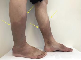
Figure 1 The patient’s lower extremities presented pretibial myxedema before therapy.
The bilateral pretibial and ankle skin was thickened with a hard texture, uneven surface, and non-pitting edema (yellow arrow).
Figure 2 Pathology of pretibial skin.
A and B: The biopsy specimens showed hyperkeratosis of squamous epithelium and perivascular inflammatory cell infiltration under hematoxylin and eosin staining; C and D: Alcian blue staining showing mucin-like substances deposited between collagens and the widened gaps of the collagenous fibers (A: HE staining, × 20; B: HE staining, × 40; C: alcian blue staining, × 20; D: alcian blue staining, × 40).
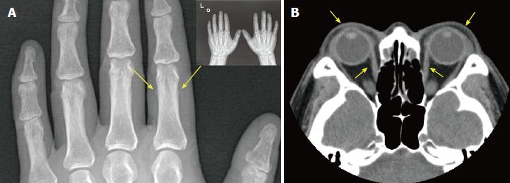
Figure 3 Imaging results of hands and eye socket.
A: X-ray scan showing the periosteal reaction in the phalangeal bone of the index finger of the left hand; B: Computed tomography scan of the eye socket showing that the bilateral extraocular muscles were slightly thickened and that both eyeballs extruded slightly.
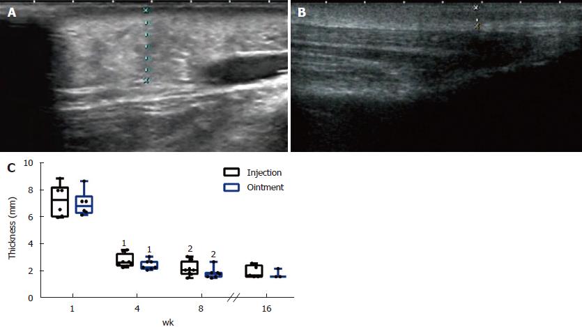
Figure 4 Ultrasound assessment of the pretibial skin area in pretibial myxedema.
A: The pretibial skin of the patient; B: The pretibial skin of the patient at 4 mo after the first injection. The pretibial skin of the patient at baseline (A) was obviously thicker than that at 4 mo after the first injection (B); C: The comparison of skin lesion thickness. 1Compared with baseline in the same leg; 2Compared with 4 wk after treatment in the same leg.
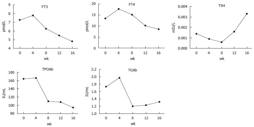
Figure 5 Increase in the levels of FT3, FT4, TPOAb, and TgAb and decrease in TSH during the first month after treatment.
Thereafter, FT3, FT4, TPOAb, and TgAb declined and TSH levels rose gradually.
Figure 6 The contrast in the presentation of pretibial myxedema before and after glucocorticoid therapy.
A: Pretibial myxedema at baseline; B: Four months after the initiation of local glucocorticoid treatment; C: Seven months after the end of treatment. Compared with baseline (A), pretibial myxedema remitted completely 4 mo after the initiation of local glucocorticoid treatment (B). Seven months after the end of treatment, no recurrence of pretibial myxedema was observed (C).
- Citation: Zhang F, Lin XY, Chen J, Peng SQ, Shan ZY, Teng WP, Yu XH. Intralesional and topical glucocorticoids for pretibial myxedema: A case report and review of literature. World J Clin Cases 2018; 6(14): 854-861
- URL: https://www.wjgnet.com/2307-8960/full/v6/i14/854.htm
- DOI: https://dx.doi.org/10.12998/wjcc.v6.i14.854


