Copyright
©The Author(s) 2018.
World J Clin Cases. Nov 26, 2018; 6(14): 842-846
Published online Nov 26, 2018. doi: 10.12998/wjcc.v6.i14.842
Published online Nov 26, 2018. doi: 10.12998/wjcc.v6.i14.842
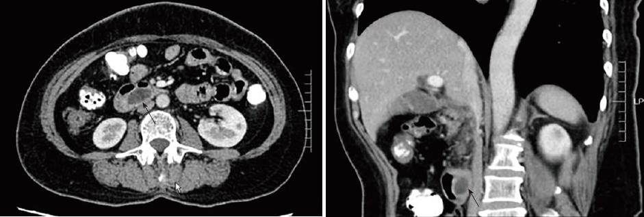
Figure 1 Multislice spiral computed tomography indicating a cystic lesion at the duodenum.
Black arrows indicate the cystic lesion.
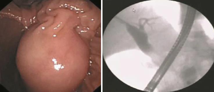
Figure 2 Duodenoscopy showing a huge submucosal mass connected to the major duodenal papilla.
The distal segment of the common bile duct was not evident.
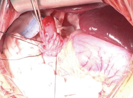
Figure 3 The cyst lesion was found to be attached to the duodenal papilla during the surgery.
- Citation: Yang J, Xiao GF, Li YX. Open surgical treatment of choledochocele: A case report and review of literature. World J Clin Cases 2018; 6(14): 842-846
- URL: https://www.wjgnet.com/2307-8960/full/v6/i14/842.htm
- DOI: https://dx.doi.org/10.12998/wjcc.v6.i14.842


