Copyright
©The Author(s) 2018.
World J Clin Cases. Nov 26, 2018; 6(14): 791-799
Published online Nov 26, 2018. doi: 10.12998/wjcc.v6.i14.791
Published online Nov 26, 2018. doi: 10.12998/wjcc.v6.i14.791
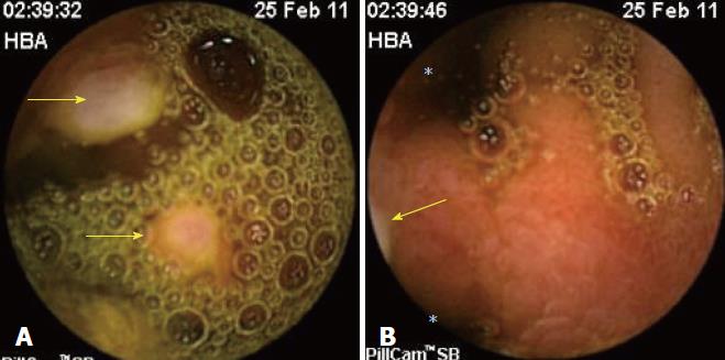
Figure 1 Image of ileum with video capsule endoscopy.
Presence of double lumen is shown (asterisks), with one having a circumferential ulcer with irregular border (arrow).
Figure 2 Image of ileum with video capsule endoscopy.
A: Ulcerated polyps in the intestinal lumen (arrows); B: Double lumen (asterisks), with the lower lumen containing the polyps (arrow).
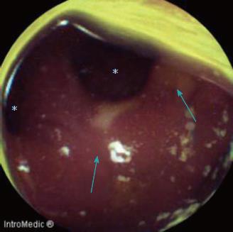
Figure 3 Image of ileum with video capsule endoscopy.
Double lumen (asterisks) with ulcerations in the diverticulum (arrows).
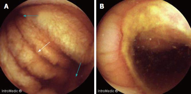
Figure 4 Image of ileum with video capsule endoscopy.
A: Double lumen (blue arrows) and diaphragm (white arrow); B: Severe circumferential ulcer in one of the lumens.
Figure 5 Image of ileum with video capsule endoscopy.
A: Polypoid image given by everted Meckelâs diverticulum; B: True polyps (arrow) inside the Meckelâs diverticulum.
- Citation: García-Compeán D, Jiménez-Rodríguez AR, Del Cueto-Aguilera ÁN, Herrera-Quiñones G, González-González JA, Maldonado-Garza HJ. Meckel’s diverticulum diagnosis by video capsule endoscopy: A case report and review of literature. World J Clin Cases 2018; 6(14): 791-799
- URL: https://www.wjgnet.com/2307-8960/full/v6/i14/791.htm
- DOI: https://dx.doi.org/10.12998/wjcc.v6.i14.791


