Copyright
©The Author(s) 2018.
World J Clin Cases. Nov 26, 2018; 6(14): 781-785
Published online Nov 26, 2018. doi: 10.12998/wjcc.v6.i14.781
Published online Nov 26, 2018. doi: 10.12998/wjcc.v6.i14.781
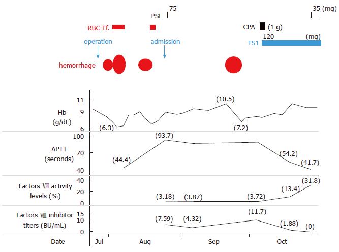
Figure 1 Clinical course in case 1.
RBC-Tf: Red blood cell transfusion; Hb: Hemoglobin; APTT: Activated partial thromboplastin time; PSL: Prednisone; CPA: Cyclophosphamide; TS1: Tegafur/gimeracil/oteracil.
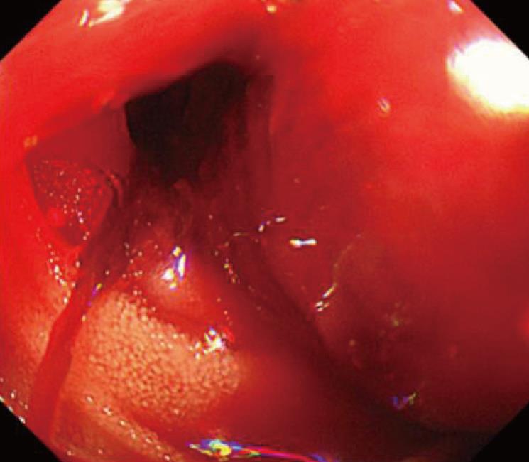
Figure 2 Esophagogastroduodenoscopy imaging (Case 1).
Intestinal bleeding was detected from the site of anastomosis.
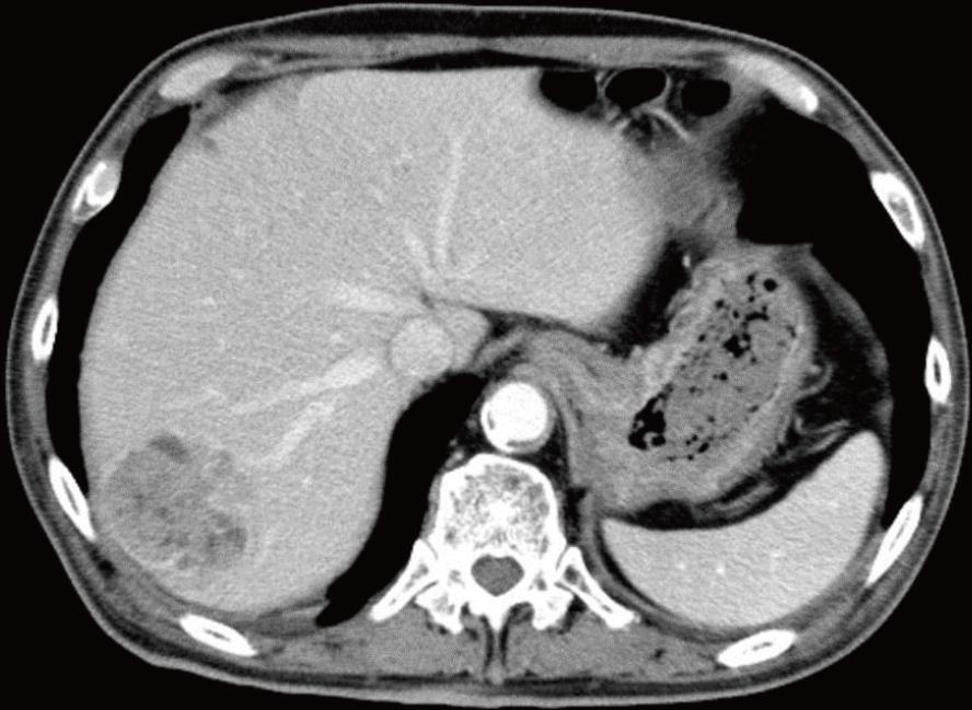
Figure 3 Abdominal computed tomography imaging (Case 2).
HCC (5.5 cm in diameter) was noted in segment 7 of the liver.
- Citation: Saito M, Ogasawara R, Izumiyama K, Mori A, Kondo T, Tanaka M, Morioka M, Ieko M. Acquired hemophilia A in solid cancer: Two case reports and review of the literature. World J Clin Cases 2018; 6(14): 781-785
- URL: https://www.wjgnet.com/2307-8960/full/v6/i14/781.htm
- DOI: https://dx.doi.org/10.12998/wjcc.v6.i14.781


