Copyright
©The Author(s) 2018.
World J Clin Cases. Nov 26, 2018; 6(14): 759-766
Published online Nov 26, 2018. doi: 10.12998/wjcc.v6.i14.759
Published online Nov 26, 2018. doi: 10.12998/wjcc.v6.i14.759
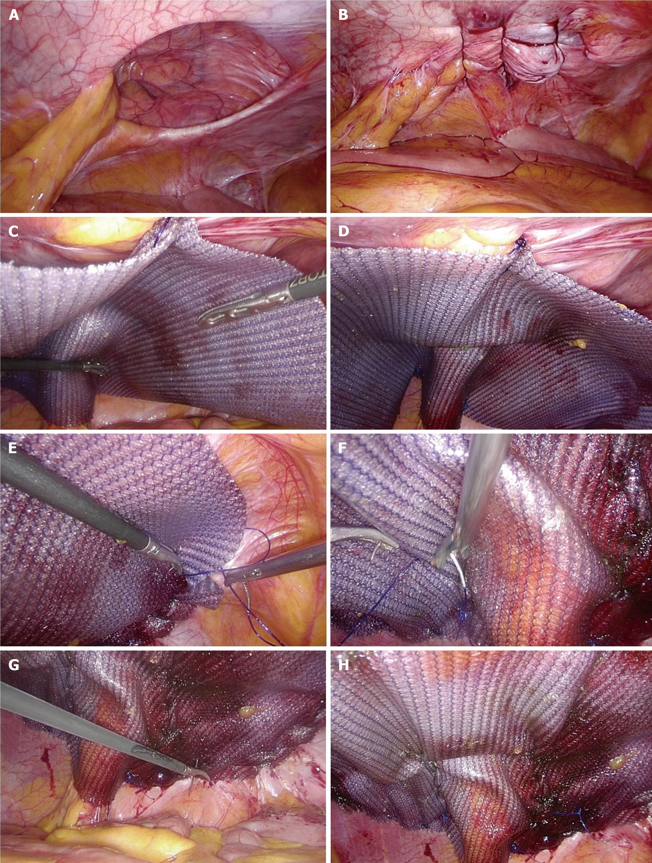
Figure 1 Keynotes for the surgical technique.
A: The parastomal defect is exposed after adhesiolysis; B: The defect was closed by interrupted stitches under laparoscopy; C: The mesh was fixed at the first point, which also covers the ostomic intestine; D: Completion of three-point anchoring; E: The mesh was fixed by continuous suturing; F: The funnel was constructed by continuous suturing; G: The transfascial suture with a purse-string needle; H: The mesh was finally fixed to the peritoneal wall.
- Citation: Huang DY, Pan L, Chen QL, Cai XY, Fang J. Modified laparoscopic Sugarbaker repair of parastomal hernia with a three-point anchoring technique. World J Clin Cases 2018; 6(14): 759-766
- URL: https://www.wjgnet.com/2307-8960/full/v6/i14/759.htm
- DOI: https://dx.doi.org/10.12998/wjcc.v6.i14.759


