Copyright
©The Author(s) 2018.
World J Clin Cases. Nov 6, 2018; 6(13): 679-682
Published online Nov 6, 2018. doi: 10.12998/wjcc.v6.i13.679
Published online Nov 6, 2018. doi: 10.12998/wjcc.v6.i13.679
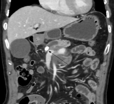
Figure 1 Computed tomography scan of the patient’s abdomen, showing chronic pancreatitis with an atrophic gland and a dilated pancreatic duct containing one stone located at the pancreas neck (indicated by an arrow).
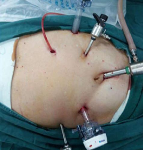
Figure 2 Intraoperative photograph showing the sites of the port wounds.
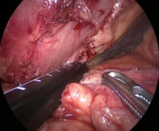
Figure 3 Laparoscopic ultrasound photograph showing the sites of the pancreatic main duct and stone.
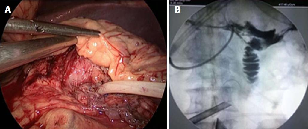
Figure 4 Intraoperative photography illustrating the insertion of one Fr14 T-type tube into the pancreatic main duct (A), it is clearly visible that the pancreatic proximal duct is unobstructed; no stone is present (B).
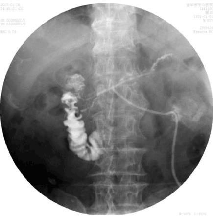
Figure 5 Postoperative X-ray radiography through the T-type tube showing that the pancreatic proximal duct is unobstructed and the absence of a stone.
- Citation: Bai Y, Yu SA, Wang LY, Gong DJ. Laparoscopic pancreatic duct incision and stone removal and T-type tube drainage for pancreatic duct stone: A case report and review of literature. World J Clin Cases 2018; 6(13): 679-682
- URL: https://www.wjgnet.com/2307-8960/full/v6/i13/679.htm
- DOI: https://dx.doi.org/10.12998/wjcc.v6.i13.679


