Copyright
©The Author(s) 2018.
World J Clin Cases. Nov 6, 2018; 6(13): 641-649
Published online Nov 6, 2018. doi: 10.12998/wjcc.v6.i13.641
Published online Nov 6, 2018. doi: 10.12998/wjcc.v6.i13.641
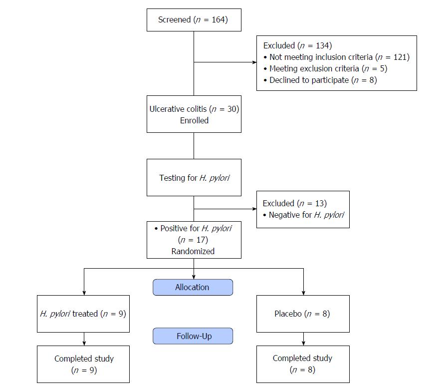
Figure 1 Study analysis population.
H. pylori: Helicobacter pylori.
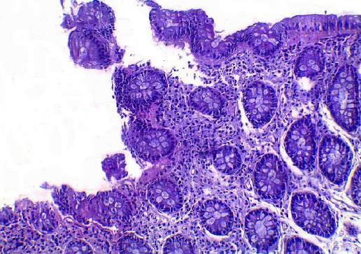
Figure 2 Demonstrates diffuse mononuclear inflammatory infiltrate in lamina propria and neutrophilic infiltration of the intestinal mucosa with ulceration of the surface epithelium as early changes of ulcerative colitis (HE: × 200).
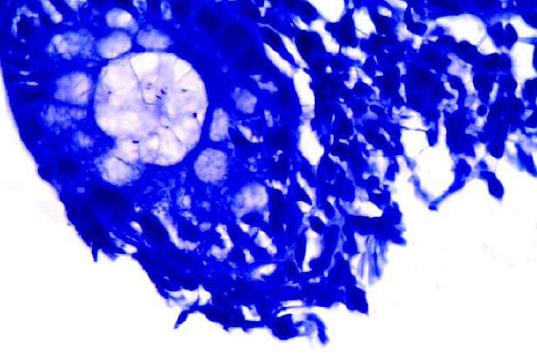
Figure 3 Giemsa staining of the previous case showing Helicobacter pylori positive rod shape organisms (Giemsa stain: × 1000).
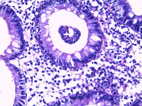
Figure 4 Ulcerative colitis showing crypt abscess and diffuse mononuclear inflammatory infiltrate in lamina propria with eosinophilia (HE: × 400).
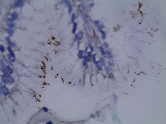
Figure 5 Immunoperoxidase staining showing Helicobacter pylori positive organisms stained brown in color (immunoperoxidase stain: × 1000).
- Citation: Mansour L, El-Kalla F, Kobtan A, Abd-Elsalam S, Yousef M, Soliman S, Ali LA, Elkhalawany W, Amer I, Harras H, Hagras MM, Elhendawy M. Helicobacter pylori may be an initiating factor in newly diagnosed ulcerative colitis patients: A pilot study. World J Clin Cases 2018; 6(13): 641-649
- URL: https://www.wjgnet.com/2307-8960/full/v6/i13/641.htm
- DOI: https://dx.doi.org/10.12998/wjcc.v6.i13.641


