Copyright
©The Author(s) 2018.
World J Clin Cases. Oct 26, 2018; 6(12): 564-569
Published online Oct 26, 2018. doi: 10.12998/wjcc.v6.i12.564
Published online Oct 26, 2018. doi: 10.12998/wjcc.v6.i12.564
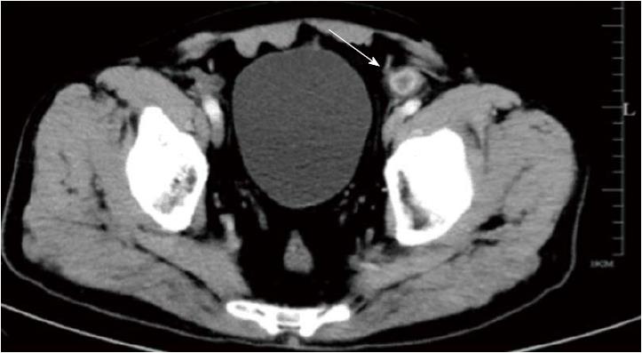
Figure 1 Abdominal computed tomography findings.
Abdominal computed tomography showed bowel wall thickening and inflammatory stranding involving the colosigmoid junction (white arrow).
Figure 2 Endoscopic findings.
A: Colonoscopy revealed a polypoid lesion in the sigmoid colon, which was hyperemic and oozed a pus-like substance; B: Endoscopic ultrasonography showed a mucosal lesion (1.23 cm × 0.62 cm) with a cavity-like structure below in sectional dimension.
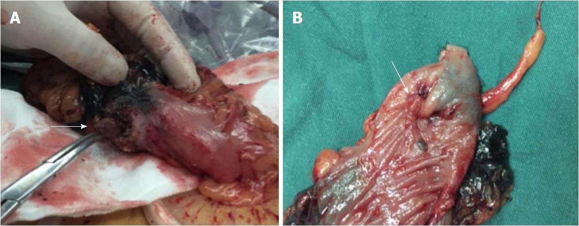
Figure 3 Intraoperative findings.
A: Mesh (arrow) penetrated the sigmoid colon and was intimately involved in the bowel wall; B: The “polyp” (slanted arrow) was observed on the luminal side of the bowel wall.
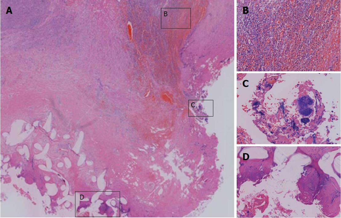
Figure 4 Histological findings revealed by Hematoxylin and Eosin staining of paraffin-embedded sections from the surgical specimen.
A: The presence of a foreign body in the bowel wall, which caused inflammatory infiltrate and granulation tissue formation in the surrounding tissue (magnification × 10); B: Infiltration of massive inflammatory cells and formation of granulation tissue (magnification × 100); C: Foreign-body giant cells were observed (magnification × 200); D: Prosthetic mesh material (magnification × 100).
- Citation: Liu S, Zhou XX, Li L, Yu MS, Zhang H, Zhong WX, Ji F. Mesh migration into the sigmoid colon after inguinal hernia repair presenting as a colonic polyp: A case report and review of literature. World J Clin Cases 2018; 6(12): 564-569
- URL: https://www.wjgnet.com/2307-8960/full/v6/i12/564.htm
- DOI: https://dx.doi.org/10.12998/wjcc.v6.i12.564


