Copyright
©The Author(s) 2018.
World J Clin Cases. Oct 26, 2018; 6(12): 538-541
Published online Oct 26, 2018. doi: 10.12998/wjcc.v6.i12.538
Published online Oct 26, 2018. doi: 10.12998/wjcc.v6.i12.538
Figure 1 Computed tomography scan - axial view.
A heterogeneously thickened bowel wall with an infiltrating tumor and separated fluid collection with air bubbles. The scan is suggestive of a perforated sigmoid tumor and an adjacent intraperitoneal abscess.
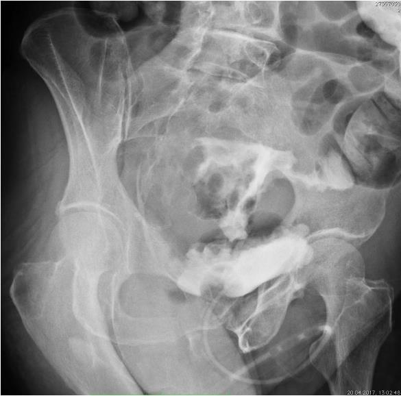
Figure 2 Cystography - an oblique projection.
An aqueous solution of the contrast medium was administered through a Foley catheter. A poorly filled urinary bladder with an irregular outline, a thickened and trabeculated wall. The contrast escapes upward and forms an irregular shape, which is the origin of the strip that contrasts a colon loop. The X-ray is suggestive of a colovesical fistula.
- Citation: Skierucha M, Barud W, Baraniak J, Krupski W. Colovesical fistula as the initial manifestation of advanced colon cancer: A case report and review of literature. World J Clin Cases 2018; 6(12): 538-541
- URL: https://www.wjgnet.com/2307-8960/full/v6/i12/538.htm
- DOI: https://dx.doi.org/10.12998/wjcc.v6.i12.538


