Copyright
©The Author(s) 2018.
World J Clin Cases. Oct 6, 2018; 6(11): 455-458
Published online Oct 6, 2018. doi: 10.12998/wjcc.v6.i11.455
Published online Oct 6, 2018. doi: 10.12998/wjcc.v6.i11.455
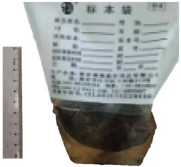
Figure 1 Photograph of the lump.
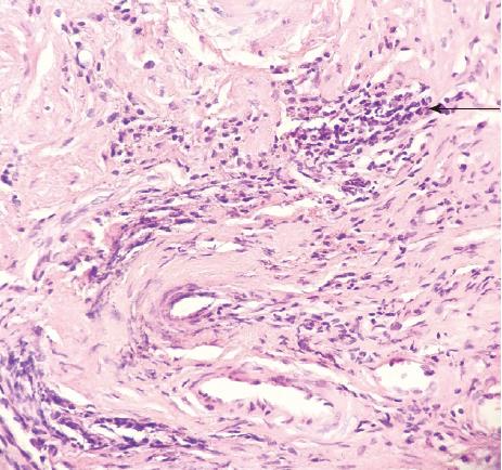
Figure 2 Proliferation of spindle cells in the lump (HE, × 200).
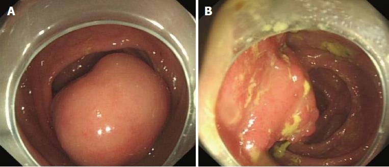
Figure 3 Endoscopic images of isolated nodularity of the ileocecus (A) and polyps of sigmoid colon (B).
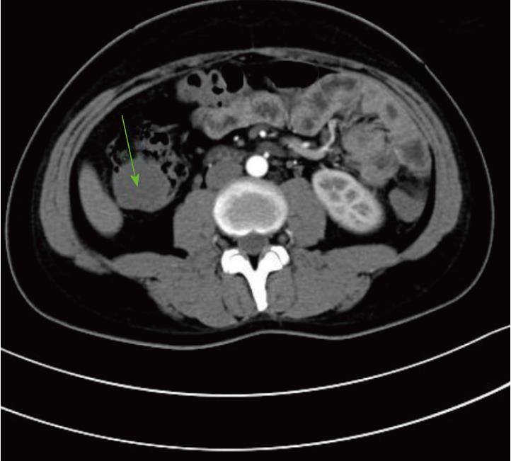
Figure 4 Photograph of computed tomography-scan abdomen shows hypoattenuating tumor of the ascending colon (green arrow).
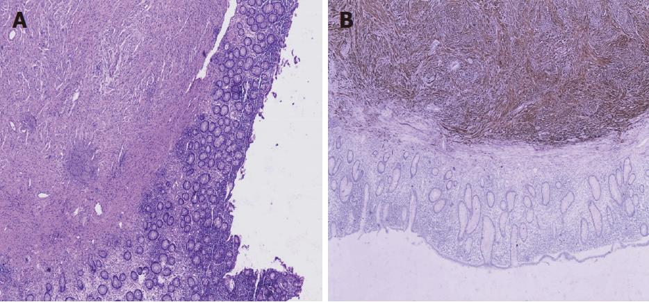
Figure 5 Photograph of resected colon (HE, × 200) (A) and immunohistochemical stain for S-100 protein (× 200) (B).
- Citation: Miao Y, Wang JJ, Chen ZM, Zhu JL, Wang MB, Cai SQ. Neurofibroma discharged from the anus with stool: A case report and review of literature. World J Clin Cases 2018; 6(11): 455-458
- URL: https://www.wjgnet.com/2307-8960/full/v6/i11/455.htm
- DOI: https://dx.doi.org/10.12998/wjcc.v6.i11.455


