Copyright
©The Author(s) 2017.
World J Clin Cases. Sep 16, 2017; 5(9): 368-372
Published online Sep 16, 2017. doi: 10.12998/wjcc.v5.i9.368
Published online Sep 16, 2017. doi: 10.12998/wjcc.v5.i9.368
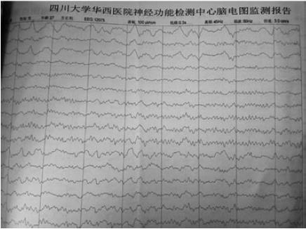
Figure 1 Electroencephalogram monitoring revealed low amplitude slow wave almost universally.
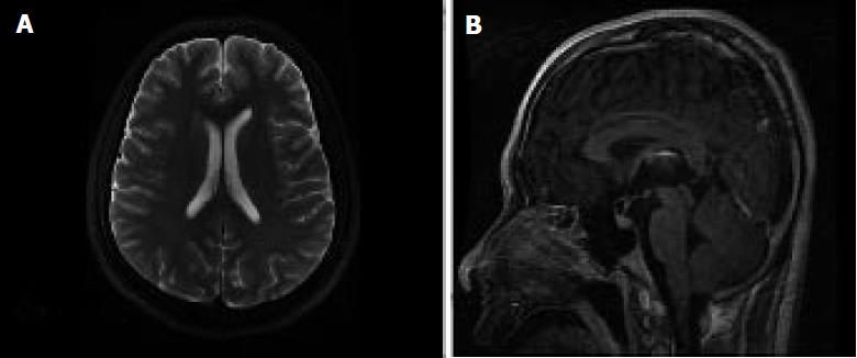
Figure 2 Transverse (A) and sagittal (B) views of magnetic resonance imaging was normal on admission.
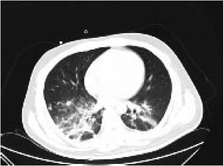
Figure 3 Computerized tomography in the chest revealed acute lung inflammation.
- Citation: Wang CC, Li DJ, Xia YQ, Liu K. Anti-N-methyl-D-aspartate receptor encephalitis that aggravates after acinetobacter baumannii pneumonia: A case report. World J Clin Cases 2017; 5(9): 368-372
- URL: https://www.wjgnet.com/2307-8960/full/v5/i9/368.htm
- DOI: https://dx.doi.org/10.12998/wjcc.v5.i9.368


