Copyright
©The Author(s) 2017.
World J Clin Cases. Sep 16, 2017; 5(9): 364-367
Published online Sep 16, 2017. doi: 10.12998/wjcc.v5.i9.364
Published online Sep 16, 2017. doi: 10.12998/wjcc.v5.i9.364
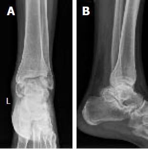
Figure 1 Anteroposterior (A) and lateral (B) radiographs of left ankle show a cystic-like lesion on posteromedial of ankle joint with destruction of articular surfaces.
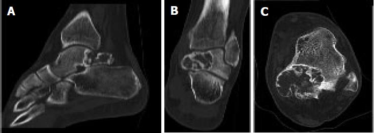
Figure 2 Sagittal (A), coronal (B), and axial (C) cuts of computed tomography scan in favor of tibiotalar and subtalar joint involvement.
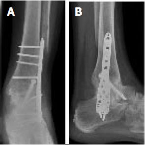
Figure 3 Anteroposterior (A) and lateral (B) radiographs of left ankle shows the bone healing without any obvious recurrence at 18 mo following surgery.
- Citation: Vosoughi AR, Mozaffarian K, Erfani MA. Recurrent aneurysmal bone cyst of talus resulted in tibiotalocalcaneal arthrodesis. World J Clin Cases 2017; 5(9): 364-367
- URL: https://www.wjgnet.com/2307-8960/full/v5/i9/364.htm
- DOI: https://dx.doi.org/10.12998/wjcc.v5.i9.364


