Copyright
©The Author(s) 2017.
World J Clin Cases. Aug 16, 2017; 5(8): 344-348
Published online Aug 16, 2017. doi: 10.12998/wjcc.v5.i8.344
Published online Aug 16, 2017. doi: 10.12998/wjcc.v5.i8.344
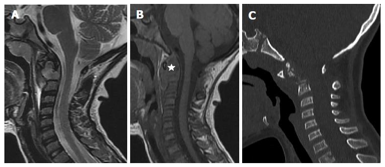
Figure 1 Initial cervical imaging: Sagittal FSET2 (A), SET1 magnetic resonance images (B) and Sagittal thin slice CT image (C).
Infiltrative mass involving the dens of C2 hypointense on T1 and hyperintense on T2 sequence, extending to the surrounding soft tissues (star) leading to an increase in C1-C2 space. No compression of the spinal cervical cord. No signal abnormality nor rupture of the posterior longitudinal ligament spine. Complement CT showed fragmented dens with important C1-C2 dislocation.
Figure 2 Langerhansien histiocytosis histology.
A: Inflammatory granuloma with esinophils and histiocytes with circonvoluted nuclei; B: Positivity of the immunostain by the antibody anti Ps100; C: Positivity of the immunostain by the antibody anti CD1a.
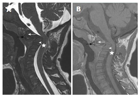
Figure 3 Six weeks follow-up after chemotherapy.
Sagittal FSET2 (A), SET1 (B) magnetic resonance images: Displaced horizontal fracture of the dens responsible for a posterior wall recoil reducing cervical occipital hinge without intramedullary signal abnormality. Increase in the shrinkage of the cervical canal despite the regression of the infiltrative process. Black arrow: Anterior arch of C1; White arrow: The upper part of dens process; White arrowhead: The base of the dens; Star: Spinal cord.
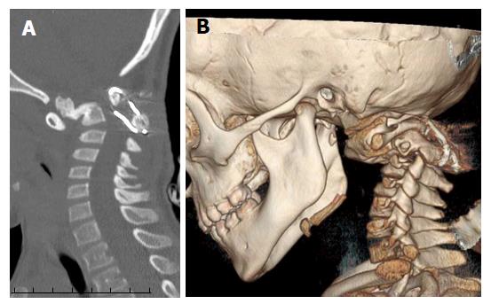
Figure 4 Five months post-operative follow-up.
Sagittal thin slice CT image (A) and volume rendering CT image (B): Consolidation of C2 fracture with moderate stenosis of the occipital hing.
- Citation: Tfifha M, Gaha M, Mama N, Yacoubi MT, Abroug S, Jemni H. Atlanto-axial langerhans cell histiocytosis in a child presented as torticollis. World J Clin Cases 2017; 5(8): 344-348
- URL: https://www.wjgnet.com/2307-8960/full/v5/i8/344.htm
- DOI: https://dx.doi.org/10.12998/wjcc.v5.i8.344


