Copyright
©The Author(s) 2017.
World J Clin Cases. Jul 16, 2017; 5(7): 292-298
Published online Jul 16, 2017. doi: 10.12998/wjcc.v5.i7.292
Published online Jul 16, 2017. doi: 10.12998/wjcc.v5.i7.292
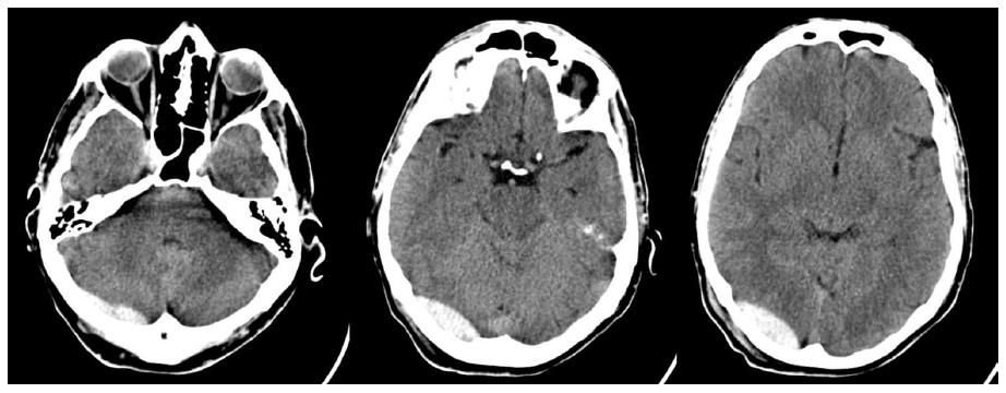
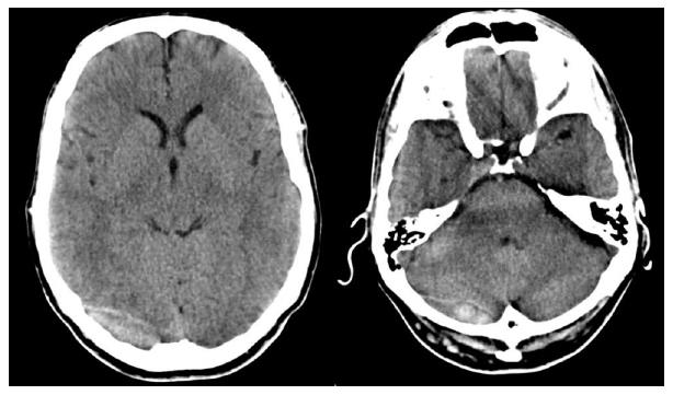
Figure 1 First computed tomography scan performed after the trauma.
It shows the presence of a combined supra-subtentorial epidural hematoma.
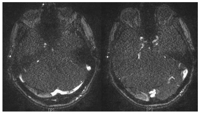
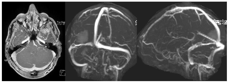
Figure 2 Angio-magnetic resonance imaging documenting the absence of the blood signal within the sinus as well as the epidural hematoma compressing the cerebellum and the sinus wall.
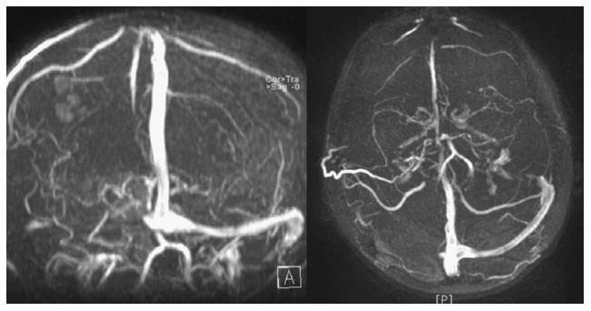
Figure 3 Angio-magnetic resonance imaging three dimensional reconstruction of the dural sinus system.
It is not possible to appreciate any signal within the right transverse sinus as it happens for dural sinus occlusion. Notice the patency of the contralateral dural sinus complex.
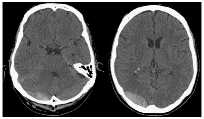
Figure 4 Computed tomography scan performed 48 h after the trauma.
An increase of the size of the ematoma was identified by the radiologist. As a consequence we decided to postpone the administration of low molecular weight heparin.
Figure 5 By the 10th post-traumatic day two more brain computed tomography scan had been performed showing the partial reabsorption of the hematoma.
From this moment the administration of low molecular weight heparin heparin began.
Figure 6 Brain magnetic resonance imaging with angiographic reconstruction of the venous system performed on 24th post-traumatic day after the onset of posterior cranial fossa symptoms.
Magnetic resonance imaging shows the partial recanalization of the right transverse sinus as well as the almost complete reabsorption of the epidural hematoma.
- Citation: Pescatori L, Tropeano MP, Mancarella C, Prizio E, Santoro G, Domenicucci M. Post traumatic dural sinus thrombosis following epidural hematoma: Literature review and case report. World J Clin Cases 2017; 5(7): 292-298
- URL: https://www.wjgnet.com/2307-8960/full/v5/i7/292.htm
- DOI: https://dx.doi.org/10.12998/wjcc.v5.i7.292


