Copyright
©The Author(s) 2017.
World J Clin Cases. Jun 16, 2017; 5(6): 254-257
Published online Jun 16, 2017. doi: 10.12998/wjcc.v5.i6.254
Published online Jun 16, 2017. doi: 10.12998/wjcc.v5.i6.254
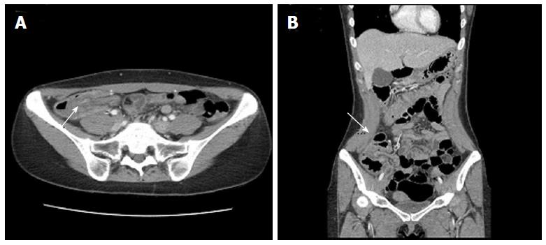
Figure 1 Axial (A) and coronal (B) view of abdominal computed tomography scans demonstrated an ileocolic intussusception with diffuse wall thickening of the ascending and transverse colon.
The entrance of the ileal segment into the colon is shown (arrow).
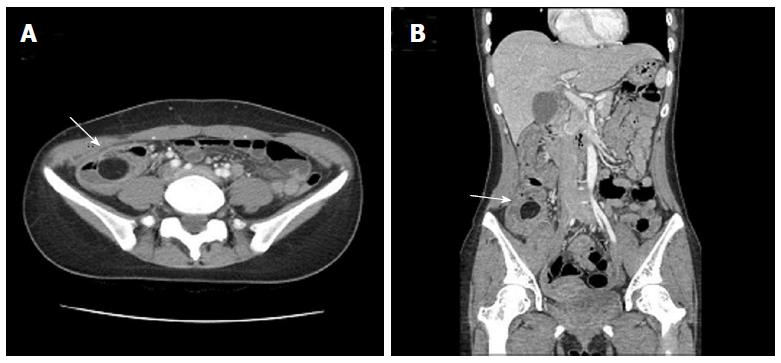
Figure 2 Axial (A) and coronal (B) plain abdominal computed tomography scans demonstrate a well-circumscribed, intraluminal hypodense mass with fat attenuation in the terminal ileum (arrow).
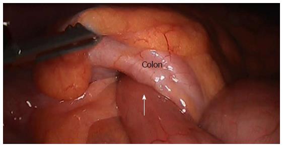
Figure 3 Laparoscopic view.
The ileum was invaginated at the colon (arrow).
- Citation: Lee DE, Choe JY. Ileocolic intussusception caused by a lipoma in an adult. World J Clin Cases 2017; 5(6): 254-257
- URL: https://www.wjgnet.com/2307-8960/full/v5/i6/254.htm
- DOI: https://dx.doi.org/10.12998/wjcc.v5.i6.254


