Copyright
©The Author(s) 2017.
World J Clin Cases. Jun 16, 2017; 5(6): 222-233
Published online Jun 16, 2017. doi: 10.12998/wjcc.v5.i6.222
Published online Jun 16, 2017. doi: 10.12998/wjcc.v5.i6.222
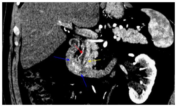
Figure 1 Preoperative computed tomography imaging.
Coronal section demonstrating a 2.1 cm × 1.4 cm periampullary duodenal mass (blue arrows). Red arrow: Common bile duct; yellow arrow: Pancreatic duct.
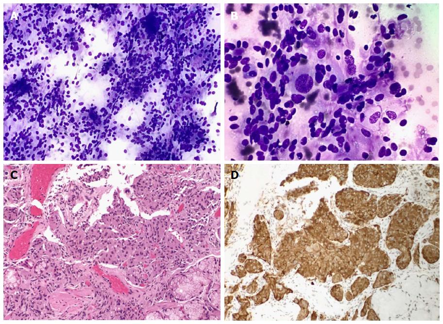
Figure 2 Fine needle aspiration biopsy and endoscopic tunneled biopsy of duodenal mass.
A: Cellular specimen with predominantly bland epithelioid cells with round to oval nuclei, Diff-Quick stain, × 20; B: Rare large ganglion-like cells with eccentric nuclei and prominent nucleoli, Diff-Quick stain, × 40; C: Epithelioid tumor cells within the duodenal lamina propria/submucosa, endoscopic tunnel biopsy, H&E stain, × 20; D: Tumor cells with positive reactivity for synaptophysin, endoscopic tunnel biopsy, × 20.
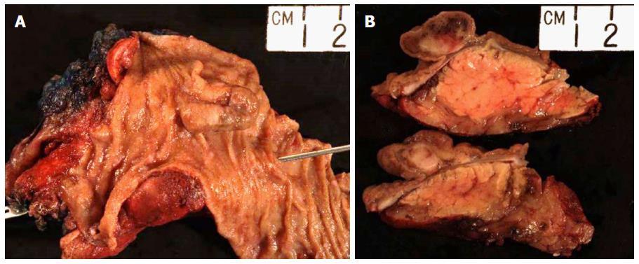
Figure 3 The tumor protruded into the duodenal lumen, 2.
0 cm proximal to the ampulla (A, probed). The mass was restricted to the duodenal submucosa, and did not invade into the adjacent pancreas (B).
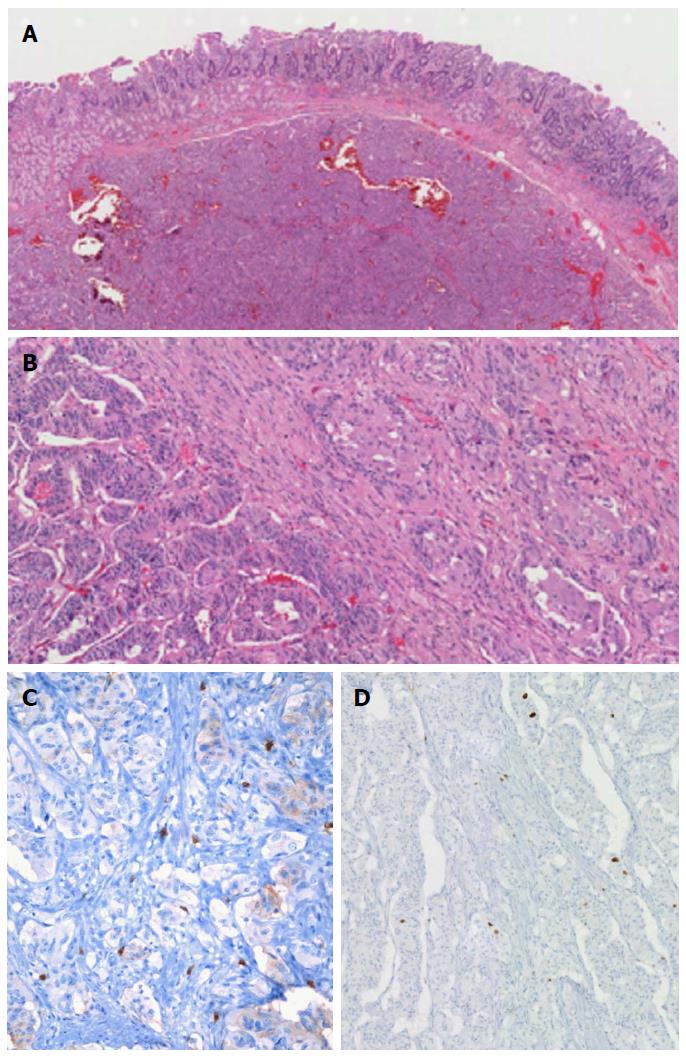
Figure 4 Primary tumor.
A: Tumor with overlying mucosa, H&E × 100; B: Tumor with epithelioid (left) and gangliocytic (right) components, H&E × 200; C: Ki-67 immunostaining proliferative index, × 100; D: CD-117 immunostaining for mast cells, × 200.
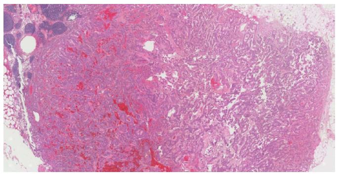
Figure 5 Lymph node metastasis, × 200.
Inset, positive immunostaining for synaptophysin, × 100.
- Citation: Cathcart SJ, Sasson AR, Kozel JA, Oliveto JM, Ly QP. Duodenal gangliocytic paraganglioma with lymph node metastases: A case report and comparative review of 31 cases. World J Clin Cases 2017; 5(6): 222-233
- URL: https://www.wjgnet.com/2307-8960/full/v5/i6/222.htm
- DOI: https://dx.doi.org/10.12998/wjcc.v5.i6.222


