Copyright
©The Author(s) 2017.
World J Clin Cases. Jun 16, 2017; 5(6): 203-211
Published online Jun 16, 2017. doi: 10.12998/wjcc.v5.i6.203
Published online Jun 16, 2017. doi: 10.12998/wjcc.v5.i6.203
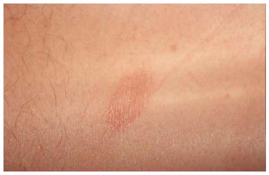
Figure 1 Herald patch.
Solitary erythemato-squamous lesion, sharply defined, round or oval, mainly located on the trunk or proximal extremities.
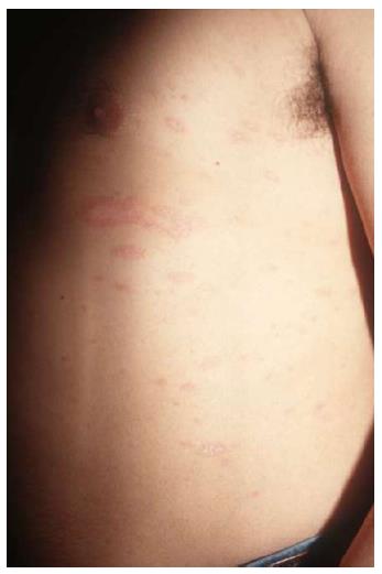
Figure 2 Classical pityriasis rosea.
Exanthematous eruption with erythemato-squamous lesions following cleavage lines on the trunk.
Figure 3 Pediatric pityriasis rosea.
Typical lesions of PR affecting an 8-mo-old boy. PR: Pityriasis rosea.
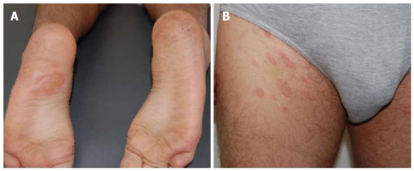
Figure 4 Herald patch in atypical location.
Herald patch on a sole (A) and (B) typical PR eruption affecting trunk and proximal thighs. PR: Pityriasis rosea.
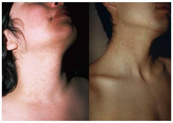
Figure 5 Inversus pityriasis rosea.
Lesions distributed on face and neck in two patients; the trunk is not affected.
Figure 6 Pityriasis rosea of the extremities.
Lesions affecting only the extremities in two different cases, without trunk involvement.
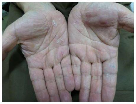
Figure 7 Acral pityriasis rosea.
Desquamation affecting the palms.
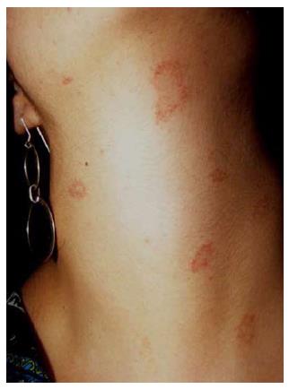
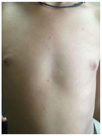
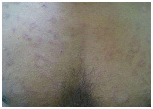
Figure 8 Purpuric pityriasis rosea.
Round and oval purpuric lesions affecting the neck of a young woman.
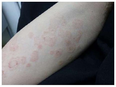
Figure 9 Urticarial pityriasis rosea.
Palpable edematous, erythematous lesions with collarette scaling.
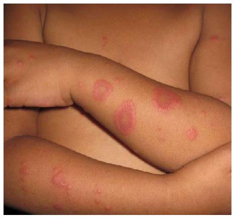
Figure 10 Erythema multiforme-like pityriasis rosea.
Annular and papular lesions resembling erythema multiforme.
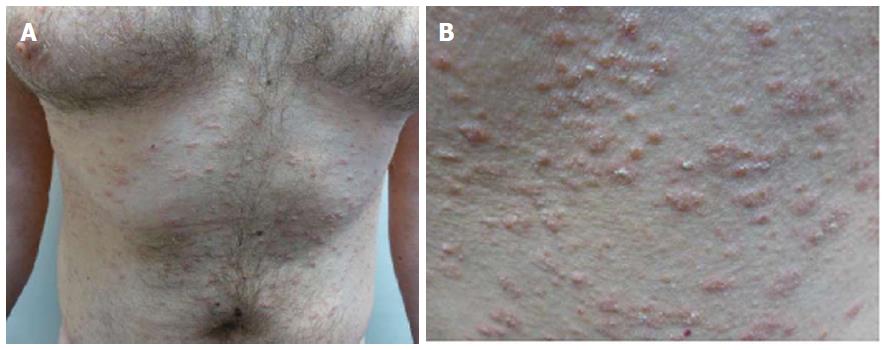
Figure 11 Papular pityriasis rosea.
A: Papular lesions with peripheral collarette (Courtesy of Priyankar Misra, Junior Resident, Dermatology, Burdwan Medical College, West Bengal, India); B: Herald patch on the neck and disseminated discrete papular eruption in a girl.
Figure 12 Follicular pityriasis rosea.
Follicular lesions with scaling (Courtesy of Shankila Mittal, Junior Resident, Dermatology, Maulana Azad Medical College, New Delhi, India).
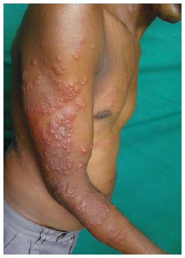
Figure 13 Vesicular pityriasis rosea.
Vesicular lesions surrounding round to oval plaques (Courtesy of Dibyendu Basu, Junior Resident, Dermatology, Medical College and Hospital, Kolkata, West Bengal, India).
Figure 14 Giant pityriasis rosea.
Large herald patch (Courtesy of Soumya Jagadeesan, Assistant Professor, Dermatology, Amrita Institute of Medical Sciences, Kochi, Kerala, India).
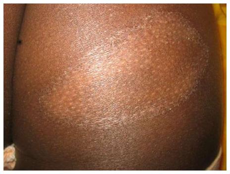
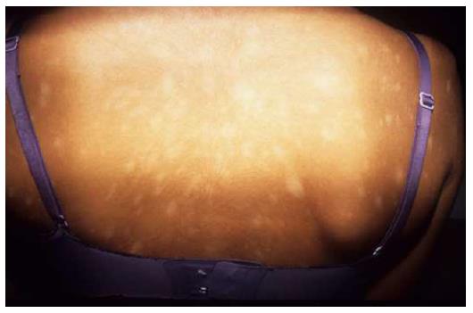
Figure 15 Hypopigmented pityriasis rosea.
Round to oval hypopigmented lesions during the whole course of the eruption.
Figure 16 Irritated pityriasis rosea.
Symptomatic eczematous lesions (Courtesy of Dipti Das, Consultant Dermatologist, Dr Marwah’s Skin Clinic, Mumbai, Maharashtra, India).
Figure 17 Pityriasis rosea-like rash.
A: The eruption in this case was probably related to the ingestion of levothyroxine in a 33-year-old man, extensively affecting the trunk; B: The lesions are small and monomorphous (Courtesy of Dr. Elizabeth Rendic).
- Citation: Urbina F, Das A, Sudy E. Clinical variants of pityriasis rosea. World J Clin Cases 2017; 5(6): 203-211
- URL: https://www.wjgnet.com/2307-8960/full/v5/i6/203.htm
- DOI: https://dx.doi.org/10.12998/wjcc.v5.i6.203


