Copyright
©The Author(s) 2017.
World J Clin Cases. Jun 16, 2017; 5(6): 191-202
Published online Jun 16, 2017. doi: 10.12998/wjcc.v5.i6.191
Published online Jun 16, 2017. doi: 10.12998/wjcc.v5.i6.191
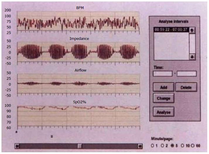
Figure 1 Cheyne-Stokes respiration in a patient with lacunar cerebral infarction[49].
BPM: Blood pressure.
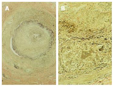
Figure 2 Temporal artery biopsy (elastin stain) in a patient (A) with Horton arteritis showing the presence of multinucleated giant cells (courtesy of Dr.
Isidro Ferrer) (B).
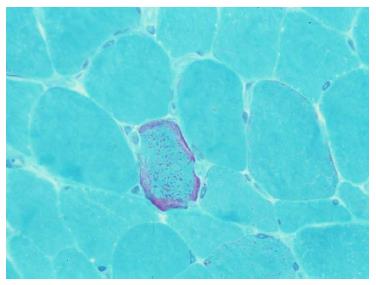
Figure 3 Ragged red fibers in muscle biopsy, stained with modified Gomori trichrome, characteristics of mitochondrial encephalomyopathy.
- Citation: Arboix A, Obach V, Sánchez MJ, Massons J. Complementary examinations other than neuroimaging and neurosonology in acute stroke. World J Clin Cases 2017; 5(6): 191-202
- URL: https://www.wjgnet.com/2307-8960/full/v5/i6/191.htm
- DOI: https://dx.doi.org/10.12998/wjcc.v5.i6.191


