Copyright
©The Author(s) 2017.
World J Clin Cases. May 16, 2017; 5(5): 183-186
Published online May 16, 2017. doi: 10.12998/wjcc.v5.i5.183
Published online May 16, 2017. doi: 10.12998/wjcc.v5.i5.183
Figure 1 The positron emission tomography-computed tomography: Left rectal wall nodule (A) and left pararectal nodule (B).
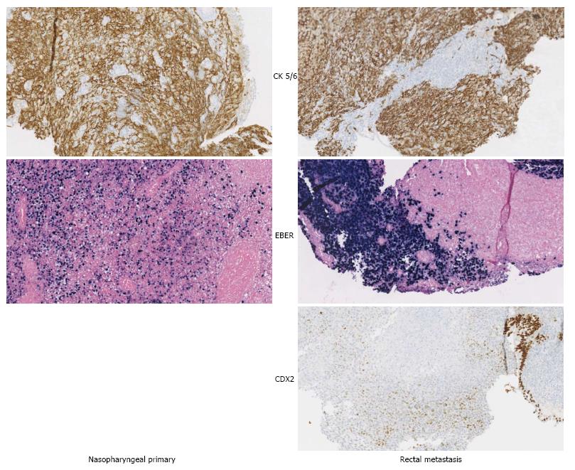
Figure 2 The nasopharyngeal primary and rectal metastasis are positive for CK 5/6 (confirming the epithelial origin) and EBER (confirming Epstein-Barr virus positivity).
CDX2 the marker of colorectal origin is negative in the rectal lesion, confirming that it is a metastasis.
- Citation: Vogel M, Kourie HR, Piccart M, Lalami Y. Unusual presentation of nasopharyngeal carcinoma with rectal metastasis. World J Clin Cases 2017; 5(5): 183-186
- URL: https://www.wjgnet.com/2307-8960/full/v5/i5/183.htm
- DOI: https://dx.doi.org/10.12998/wjcc.v5.i5.183


