Copyright
©The Author(s) 2017.
World J Clin Cases. May 16, 2017; 5(5): 178-182
Published online May 16, 2017. doi: 10.12998/wjcc.v5.i5.178
Published online May 16, 2017. doi: 10.12998/wjcc.v5.i5.178
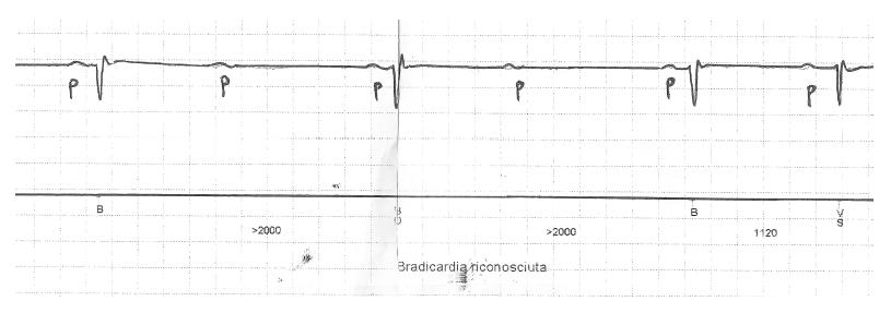
Figure 1 Patient #1.
Continuous electrocardiograph monitoring showing paroxysmal episodes of 2:1 atrioventricular block with narrow QRS and lengthening of PP interval (associated sinus bradycardia).
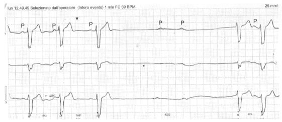
Figure 2 Patient #2.
Holter electrocardiograph monitoring showing episodes of paroxysmal complete atrioventricular block associated with PP interval lengthening. Baseline wide QRS complex.
- Citation: De Maria E, Borghi A, Modonesi L, Cappelli S. Ticagrelor therapy and atrioventricular block: Do we need to worry? World J Clin Cases 2017; 5(5): 178-182
- URL: https://www.wjgnet.com/2307-8960/full/v5/i5/178.htm
- DOI: https://dx.doi.org/10.12998/wjcc.v5.i5.178


