Copyright
©The Author(s) 2017.
World J Clin Cases. May 16, 2017; 5(5): 172-177
Published online May 16, 2017. doi: 10.12998/wjcc.v5.i5.172
Published online May 16, 2017. doi: 10.12998/wjcc.v5.i5.172
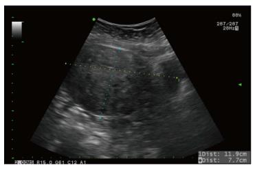
Figure 1 Ultrasound of adenomyosis of uterus.
The largest diameter of uterus measured was 11.9 cm.
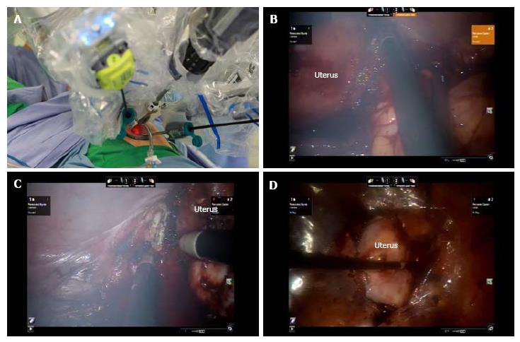
Figure 2 Intraoperative view of supracervical hysterectomy.
A: Placement of robotic trocars using a single-site device; B: Cutting right cervical region; C: Cutting left cervical region; D: Amputated uterus placed into tissue bag.
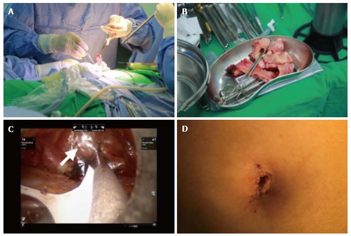
Figure 3 Intraoperative view of manual morcellation of the uterus and the placement of seprafilm.
A: Manual morcellation of uterus through the single-site wound; B: Morcellated uterus; C: Seprafilm placed onto surgical sites (arrow); D: Postoperative umbilical scar.
- Citation: Ding DC, Hong MK, Chu TY, Chang YH, Liu HW. Robotic single-site supracervical hysterectomy with manual morcellation: Preliminary experience. World J Clin Cases 2017; 5(5): 172-177
- URL: https://www.wjgnet.com/2307-8960/full/v5/i5/172.htm
- DOI: https://dx.doi.org/10.12998/wjcc.v5.i5.172


