Copyright
©The Author(s) 2017.
World J Clin Cases. Mar 16, 2017; 5(3): 112-118
Published online Mar 16, 2017. doi: 10.12998/wjcc.v5.i3.112
Published online Mar 16, 2017. doi: 10.12998/wjcc.v5.i3.112
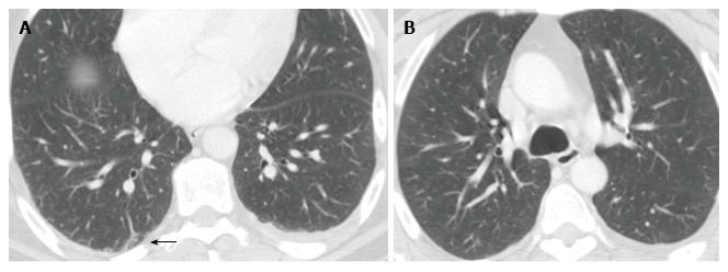
Figure 1 Chest computed tomography showing pulmonary lesions in the posterior region of the right lung (A) associated with left pulmonary embolism (B).
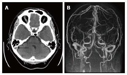
Figure 2 Internal jugular vein thrombosis (arrow) showed by brain TC (A), and further complete re-canalization demonstrated by cerebral magnetic resonance angiogram after antibiotic and anticoagulant therapy (B).
- Citation: De Giorgi A, Fabbian F, Molino C, Misurati E, Tiseo R, Parisi C, Boari B, Manfredini R. Pulmonary embolism and internal jugular vein thrombosis as evocative clues of Lemierre’s syndrome: A case report and review of the literature. World J Clin Cases 2017; 5(3): 112-118
- URL: https://www.wjgnet.com/2307-8960/full/v5/i3/112.htm
- DOI: https://dx.doi.org/10.12998/wjcc.v5.i3.112


