Copyright
©The Author(s) 2017.
World J Clin Cases. Feb 16, 2017; 5(2): 67-72
Published online Feb 16, 2017. doi: 10.12998/wjcc.v5.i2.67
Published online Feb 16, 2017. doi: 10.12998/wjcc.v5.i2.67
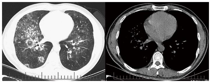
Figure 1 Pulmonary computed tomography images showing bilateral consolidation with air-bronchogram, especially in the right lung with multiple ground glass low-density shadows.
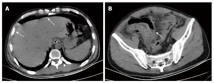
Figure 2 Abdominal transverse computed tomography images.
A: Free air in the abdomen and fluid around the liver; B: Intestinal wall thickening in the right lower quadrant and seepage, scattered with free air.
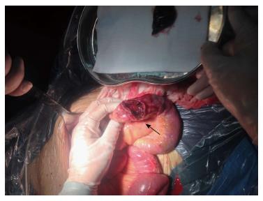
Figure 3 Introperative findings.
There were 11 perforations in the intestine. The arrow points to the bigger one measuring about 3 cm × 3 cm.
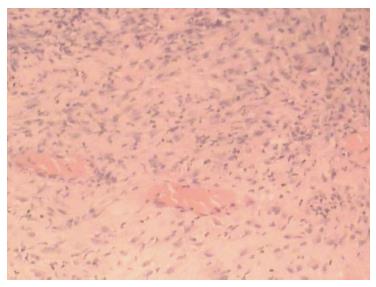
Figure 4 Histopathological examination showing a large number of infiltrated inflammatory cells (HE staining, × 100).
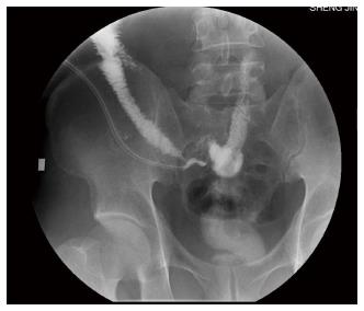
Figure 5 Fistulography showing that after injection of contrast agents, the intestine of the right lower quadrant abdomen developed a fistula.
- Citation: He JN, Tian Z, Yao X, Li HY, Yu Y, Liu Y, Liu JG. Multiple perforations and fistula formation following corticosteroid administration: A case report. World J Clin Cases 2017; 5(2): 67-72
- URL: https://www.wjgnet.com/2307-8960/full/v5/i2/67.htm
- DOI: https://dx.doi.org/10.12998/wjcc.v5.i2.67


