Copyright
©The Author(s) 2017.
World J Clin Cases. Feb 16, 2017; 5(2): 40-45
Published online Feb 16, 2017. doi: 10.12998/wjcc.v5.i2.40
Published online Feb 16, 2017. doi: 10.12998/wjcc.v5.i2.40
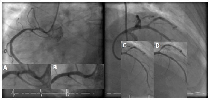
Figure 1 Initial coronary angiogram showing proximal right coronary and first diagonal stenoses.
A, B: Right coronary artery pre- and post-stenting with 3.0 Xience Everolimus drug eluting stent, respectively; C, D: Pre- and post-balloon angioplasty to first diagonal, respectively.
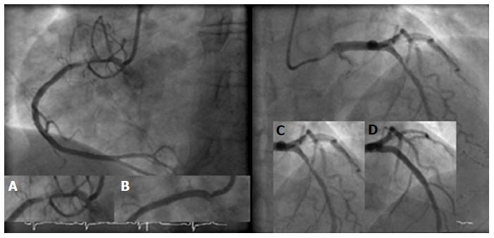
Figure 2 Second coronary angiogram following presentation with stable angina.
A, B: Severe instent restenosis in the proximal segment of RCA and result post-Paclitaxel drug eluting balloon; C, D: Severe stenosis in mid LAD segment stented and subsequently stented with 3.5 Xience Everolimus drug eluting stent. LAD: Left anterior descending artery; RCA: Right coronary artery.
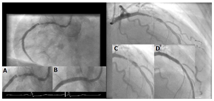
Figure 3 Coronary angiogram performed following second acute coronary syndrome event.
A, B: Severe recurrent instent restenosis within the proximal segment of RCA and subsequent Xience stent; C, D: Severe ISR within mid LAD stented segment and subsequent Xience stent. LAD: Left anterior descending artery; RCA: Right coronary artery; ISR: In stent restenosis.
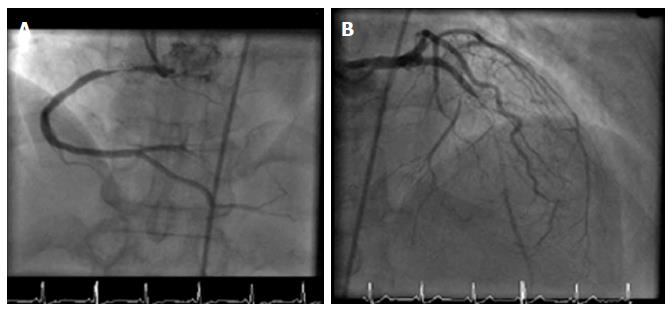
Figure 4 Further coronary angiogram following intractable angina symptoms.
A: Sub totally occluded proximal RCA within stented segment; B: Sub totally occluded LAD with antegrade filling. LAD: Left anterior descending artery; RCA: Right coronary artery.
- Citation: Alkhalil M, Conlon CP, Ashrafian H, Choudhury RP. Aggressive restenosis after percutaneous intervention in two coronary loci in a patient with human immunodeficiency virus infection. World J Clin Cases 2017; 5(2): 40-45
- URL: https://www.wjgnet.com/2307-8960/full/v5/i2/40.htm
- DOI: https://dx.doi.org/10.12998/wjcc.v5.i2.40


