Copyright
©The Author(s) 2017.
World J Clin Cases. Dec 16, 2017; 5(12): 440-445
Published online Dec 16, 2017. doi: 10.12998/wjcc.v5.i12.440
Published online Dec 16, 2017. doi: 10.12998/wjcc.v5.i12.440
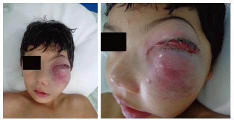
Figure 1 Initial clinical features of the lesion showing a reddish painful firm mass on left side of face with rapid evolution (25 d).
This lesion was causing left visual impairment with notorious swelling on facial skin with absence of other obstructive symptoms.
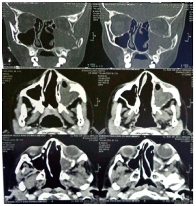
Figure 2 Computed tomography scan of the paranasal sinuses.
On coronal view, a diffuse hypodense mass was dislocating lateral wall of left sinus and compressing the inferior border of left orbital structure with tumor invasion. On axial plan, tumor mass was filling the left sinus and a dislocated nasal septum was evident.
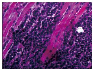
Figure 3 The microscopic slide showed an undifferentiated malignancy with hyperchromatic rounded cells with scarce and eosinophilic cytoplasma infiltrating the skeletal muscle tissue (hematoxylin-eosin, 40 ×).
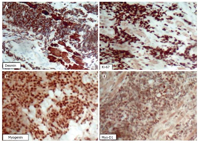
Figure 4 An immunohistochemical analysis was performed on a biopsied tumour fragment from the left maxillary sinus.
A: Immunohistochemical analysis showed positiveness to anti-Desmin antibody with dual cytoplasmatic and nuclear staining; B: The same pattern was observed against anti-Ki67 (B) showing intense positiveness and high rate of cell proliferation; C and D: Anti-myogenin and MYO-D1 were positively found on nuclear staining leading to RMS lineage supposition.
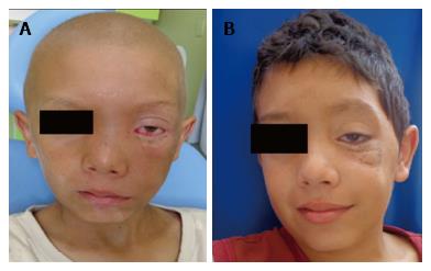
Figure 5 Monitoring of the patient after the period of 2.
5 mo of chemotherapy and after completion of treatment (18 mo). A: Monitoring of the patient after the period of 2.5 mo of chemotherapy; B: Monitoring of the patient after completion of treatment (18 mo).
- Citation: de Melo ACR, Lyra TC, Ribeiro ILA, da Paz AR, Bonan PRF, de Castro RD, Valença AMG. Embryonal rhabdomyosarcoma in the maxillary sinus with orbital involvement in a pediatric patient: Case report. World J Clin Cases 2017; 5(12): 440-445
- URL: https://www.wjgnet.com/2307-8960/full/v5/i12/440.htm
- DOI: https://dx.doi.org/10.12998/wjcc.v5.i12.440


