Copyright
©The Author(s) 2016.
World J Clinical Cases. Sep 16, 2016; 4(9): 285-289
Published online Sep 16, 2016. doi: 10.12998/wjcc.v4.i9.285
Published online Sep 16, 2016. doi: 10.12998/wjcc.v4.i9.285
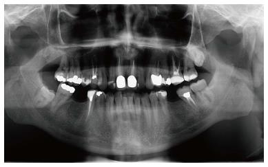
Figure 1 Pretreatment orthopantomography.
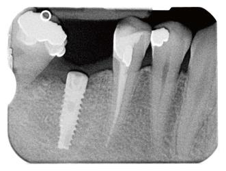
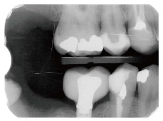
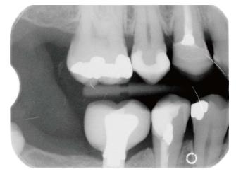
Figure 2 Lower second molar mesial angular periodontal defect.
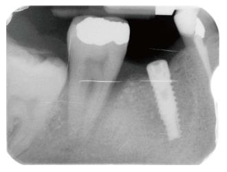
Figure 3 Worsening of lower second molar mesial angular periodontal defect.
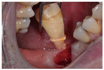
Figure 4 Lower second molar extraction.
Figure 5 Post-treatment radiograph after second molar extraction.
Figure 6 Three-year follow-up.
- Citation: Dianiskova S, Calzolari C, Migliorati M, Silvestrini-Biavati A, Isola G, Savoldi F, Dalessandri D, Paganelli C. Tooth loss caused by displaced elastic during simple preprosthetic orthodontic treatment. World J Clinical Cases 2016; 4(9): 285-289
- URL: https://www.wjgnet.com/2307-8960/full/v4/i9/285.htm
- DOI: https://dx.doi.org/10.12998/wjcc.v4.i9.285


