Copyright
©The Author(s) 2016.
World J Clinical Cases. Sep 16, 2016; 4(9): 269-272
Published online Sep 16, 2016. doi: 10.12998/wjcc.v4.i9.269
Published online Sep 16, 2016. doi: 10.12998/wjcc.v4.i9.269
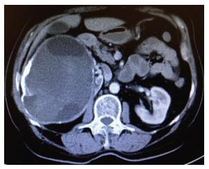
Figure 1 Contrast-enhanced abdominal computed tomography revealed a 150 mm × 120 mm mass with peripheral calcification associated with multicentric hypodense cystic necrotic areas, originating from the right adrenal gland.
- Citation: Akbulut S. Incidentally detected hydatid cyst of the adrenal gland: A case report. World J Clinical Cases 2016; 4(9): 269-272
- URL: https://www.wjgnet.com/2307-8960/full/v4/i9/269.htm
- DOI: https://dx.doi.org/10.12998/wjcc.v4.i9.269


