Copyright
©The Author(s) 2016.
World J Clinical Cases. Sep 16, 2016; 4(9): 264-268
Published online Sep 16, 2016. doi: 10.12998/wjcc.v4.i9.264
Published online Sep 16, 2016. doi: 10.12998/wjcc.v4.i9.264
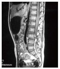
Figure 1 Initial magnetic resonance imaging, sagittal view T1-weighted, showing Chance fracture with dislocation of the 3rd lumbar vertebra and neurological compression.
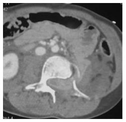
Figure 2 Initial axial computed tomographic scan showing entrapment of small bowel loop in the spinal canal.
No air was seen in the spinal canal.
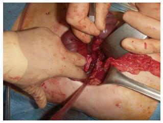
Figure 3 Intra-operative view of the jejunum loop during the second time surgical procedure.
Due to the necrotic aspect of the bowel, resection and anastomosis was performed.
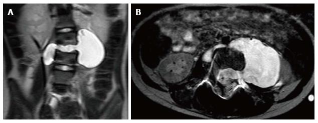
Figure 4 T2-weighted coronal (A) and axial (B) magnetic resonance imaging one month after the trauma showing LCS leakage in the abdominal cavity.
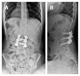
Figure 5 AP (A) and lateral (B) X-rays at two years follow-up showing L2-L4 vertebral fusion after posterior and anterior procedures.
- Citation: Pesenti S, Blondel B, Faure A, Peltier E, Launay F, Jouve JL. Small bowel entrapment and ureteropelvic junction disruption associated with L3 Chance fracture-dislocation. World J Clinical Cases 2016; 4(9): 264-268
- URL: https://www.wjgnet.com/2307-8960/full/v4/i9/264.htm
- DOI: https://dx.doi.org/10.12998/wjcc.v4.i9.264


