Copyright
©The Author(s) 2016.
World J Clinical Cases. Sep 16, 2016; 4(9): 258-263
Published online Sep 16, 2016. doi: 10.12998/wjcc.v4.i9.258
Published online Sep 16, 2016. doi: 10.12998/wjcc.v4.i9.258
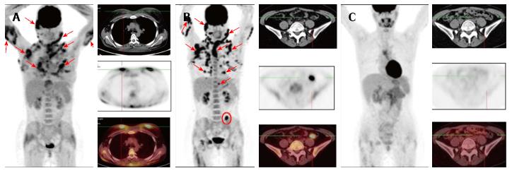
Figure 1 Case 1.
A: Before treatment, showing multiple confluent pathological foci of hypermetabolic activity in subcutaneous fat (the largest are indicated by arrows on the whole-body image) and pathological foci in the breasts (in transverse sections); B: After 6 cycles of the CHOP regimen, showing no changes in the follow-up examinations, with conservation of previously defined lesions and development of new ones in the subcutaneous tissue (indicated by arrows on the whole-body image) and pathological focus of hypermetabolic activity which had developed in the mesentery of the descending colon (indicated by circles, on the whole-body image as well); C: After 6 cycles of the GEM-P chemotherapy regimen, showing marked improvement, as indicated by resorption of all previously defined foci. CHOP: Cyclophosphamide, doxorubicine, vincristine and prednisolone; GEM-P: Gemcitabine, cisplatin and methylprednisolone.
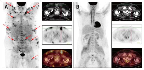
Figure 2 Case 2.
A: Before treatment, showing multiple separate and confluent pathological foci of fluorine-18 fluorodeoxyglucose uptake in local dense areas of subcutaneous fat (the largest are indicated by arrows on the whole-body image); B: After 7 cycles of the GEM-P chemotherapy regimen, showing preservation of metabolically inactive areas of consolidation of subcutaneous fat. GEM-P: Gemcitabine, cisplatin and methylprednisolone.
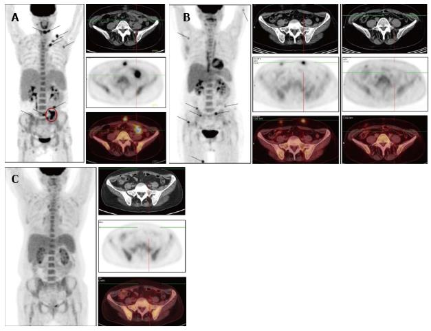
Figure 3 Case 3.
A: Before treatment, showing multiple pathological foci of hypermetabolic activity in local dense areas of subcutaneous fat (the largest are indicated by arrows, on the whole-body image as well) and pathological foci of fluorine-18 fluorodeoxyglucose uptake in the epiploon in the left mesogastric area (indicated by circles, on the whole-body image as well); B: After 3 cycles of FCM, showing resorption of all previously defined foci (including those in the greater omentum) but development of new ones in the subcutaneous tissue; C: After 3 cycles of GEM-P, showing marked improvement that was indicated by resorption of all previously defined foci. FCM: Fludarabine, mitoxantrone and cyclophosphamide; GEM-P: Gemcitabine, cisplatin and methylprednisolone.
- Citation: Gorodetskiy VR, Mukhortova OV, Aslanidis IP, Klapper W, Probatova NA. Fluorine-18 fluorodeoxyglucose positron emission tomography/computed tomography evaluation of subcutaneous panniculitis-like T cell lymphoma and treatment response. World J Clinical Cases 2016; 4(9): 258-263
- URL: https://www.wjgnet.com/2307-8960/full/v4/i9/258.htm
- DOI: https://dx.doi.org/10.12998/wjcc.v4.i9.258


