Copyright
©The Author(s) 2016.
World J Clin Cases. Aug 16, 2016; 4(8): 229-232
Published online Aug 16, 2016. doi: 10.12998/wjcc.v4.i8.229
Published online Aug 16, 2016. doi: 10.12998/wjcc.v4.i8.229
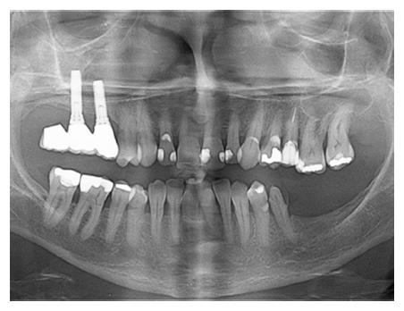
Figure 1 The preoperative panoramic radiography showed an unknown disappearance of an upper endosseous dental implant previously located in the posterior aspect of the right upper maxilla.
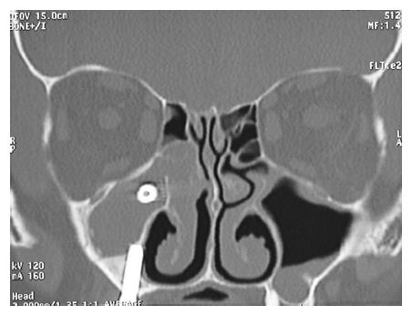
Figure 2 Coronal computed tomography scan clearly revealed the dental implant located in the upper third of the maxillary sinus.
Acute maxillary sinusitis and ethmoiditis are evident.
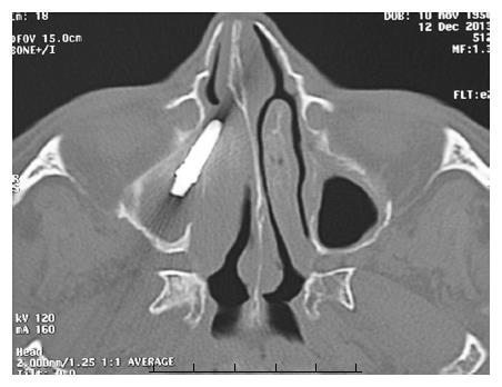
Figure 3 Axial computed tomography scan allowed to analyze the exact position of the implant between the maxillary sinus interiorly and the osteomeatal complex posteriorly.
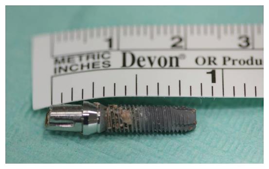
Figure 4 The sneezed implant.
The prosthetic abutment is still engaged to the dental implant.
- Citation: Procacci P, De Santis D, Bertossi D, Albanese M, Plotegher C, Zanette G, Pardo A, Nocini PF. Extraordinary sneeze: Spontaneous transmaxillary-transnasal discharge of a migrated dental implant. World J Clin Cases 2016; 4(8): 229-232
- URL: https://www.wjgnet.com/2307-8960/full/v4/i8/229.htm
- DOI: https://dx.doi.org/10.12998/wjcc.v4.i8.229


