Copyright
©The Author(s) 2016.
World J Clin Cases. Aug 16, 2016; 4(8): 202-206
Published online Aug 16, 2016. doi: 10.12998/wjcc.v4.i8.202
Published online Aug 16, 2016. doi: 10.12998/wjcc.v4.i8.202
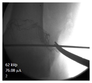
Figure 1 Start position of the guide pin, placed in the lateral femoral epicondyle.
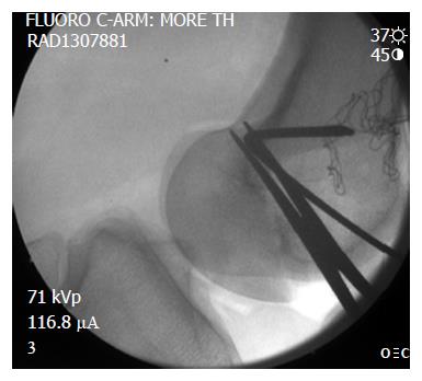
Figure 2 Position of the entrance tunnel from lateral X-ray compared to the Blumensaat line and the posterior cortex in that area.
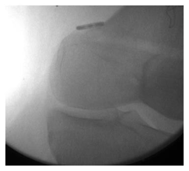
Figure 3 EndoButton type device fixed on the medial femoral condyle.
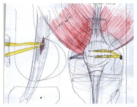
Site of graft fixation.
Figure 4 Decompression window illustration in both AP and lateral view.
- Citation: Beckert M, Crebs D, Nieto M, Gao Y, Albright J. Lateral patellofemoral ligament reconstruction to restore functional capacity in patients previously undergoing lateral retinacular release. World J Clin Cases 2016; 4(8): 202-206
- URL: https://www.wjgnet.com/2307-8960/full/v4/i8/202.htm
- DOI: https://dx.doi.org/10.12998/wjcc.v4.i8.202


