Copyright
©The Author(s) 2016.
World J Clin Cases. Jun 16, 2016; 4(6): 151-154
Published online Jun 16, 2016. doi: 10.12998/wjcc.v4.i6.151
Published online Jun 16, 2016. doi: 10.12998/wjcc.v4.i6.151
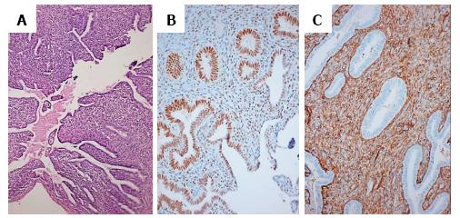
Figure 1 Endometrial tissue.
A: Endometrial tissue comprising of endometrial glands with stroma; hematoxylin and eosin stain, × 100; B: Endometrial glandular cells and stromal cells showing estrogen receptor positivity, × 100; C: Endometrial tissue showing CD10 positivity in the stromal cells, × 100.
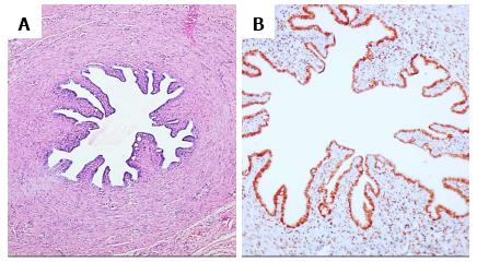
Figure 2 Fallopian tube.
A: Fallopian tube with ciliated columnar cells, hematoxylin and eosin stain, × 100; B: Fallopian tube with ciliated columnar cells, showing estrogen receptor positivity, × 100.
- Citation: Vanikar AV, Nigam LA, Patel RD, Kanodia KV, Suthar KS, Thakkar UG. Persistent mullerian duct syndrome presenting as retractile testis with hypospadias: A rare entity. World J Clin Cases 2016; 4(6): 151-154
- URL: https://www.wjgnet.com/2307-8960/full/v4/i6/151.htm
- DOI: https://dx.doi.org/10.12998/wjcc.v4.i6.151


