Copyright
©The Author(s) 2016.
World J Clin Cases. Dec 16, 2016; 4(12): 409-412
Published online Dec 16, 2016. doi: 10.12998/wjcc.v4.i12.409
Published online Dec 16, 2016. doi: 10.12998/wjcc.v4.i12.409
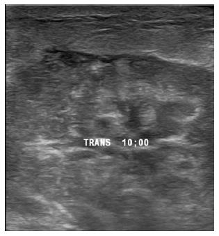
Figure 1 Ultrasound of the area of concern in the right breast reveals a large irregular hypoechoic mass without any evidence of abscess or fluid collection.
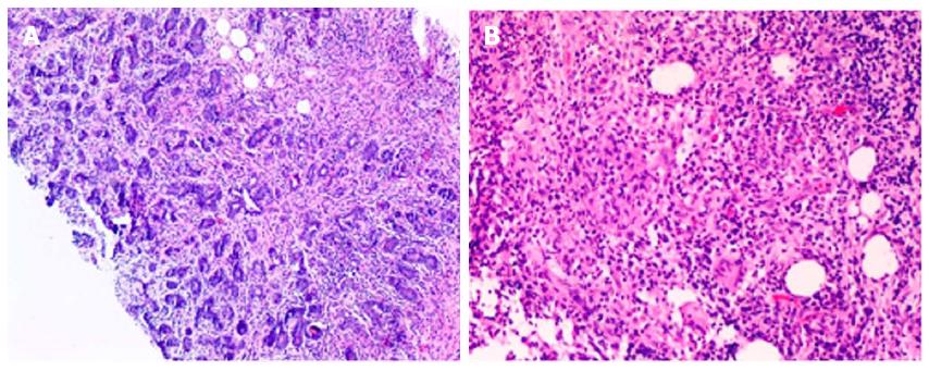
Figure 2 Core biopsy of the mass, 4 × (A) and 10 × (B) magnification, reveals granulomatous inflammatory reaction centered on lobules, with granulomas composed of epithelioid histiocytes, Landerhans giant cells accompanied by lymphocytes, plasma cells and occasional eosinophils.
- Citation: Kamyab A. Granulomatous lobular mastitis secondary to Mycobacterium fortuitum. World J Clin Cases 2016; 4(12): 409-412
- URL: https://www.wjgnet.com/2307-8960/full/v4/i12/409.htm
- DOI: https://dx.doi.org/10.12998/wjcc.v4.i12.409


