Copyright
©The Author(s) 2016.
World J Clin Cases. Nov 16, 2016; 4(11): 380-384
Published online Nov 16, 2016. doi: 10.12998/wjcc.v4.i11.380
Published online Nov 16, 2016. doi: 10.12998/wjcc.v4.i11.380
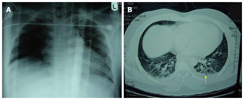
Figure 1 Antero-posterior chest X-ray performed at presentation (A) and contrast enhanced computed tomography (B).
A: Fracture right clavicle and mild haziness of the left lung field; B: Mild pleural effusion of the left side (yellow arrow).
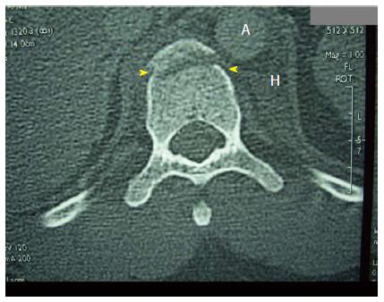
Figure 2 Computed tomography trauma shows fracture anterior rim of the body of the 10th thoracic vertebra (arrow heads), a haematoma to left of the body of the vertebra (H) and around the descending aorta (A).
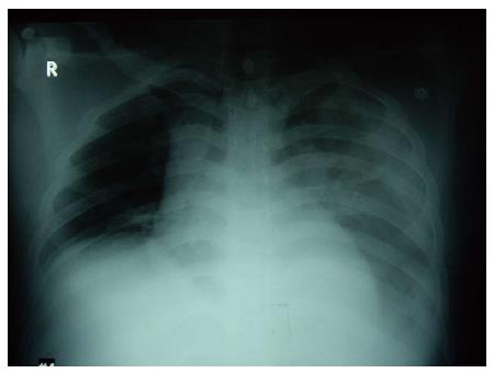
Figure 3 A portable chest X-ray performed in the intensive care unit on the second day shows whitish homogenous opacity of the left lung field.
The trachea and mediastinum are shifted to the right side.
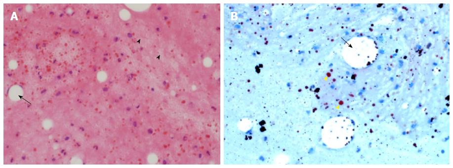
Figure 4 Histopathology (A) and Oil red O stain (B) of a cell block of the left pleural fluid.
A: Macrophages containing large fat vacuoles (black arrow) and globules of fat in the background (arrow heads). Macrophages show eccentric nuclei and “Empty looking” cytoplasm. Most of the fat has been dissolved and removed during processing, having been dissolved by the xylene and alcohol. A few mixed inflammatory cells are seen in the background (Haematoxylin and eosin, × 40); B: An Oil red O stain demonstrates fat globules of varying sizes stained orange (yellow arrow heads). Some globules are still present in the macrophages (black arrow), but most of them are extracellular, within the chylous fluid (Oil red O stain × 40).
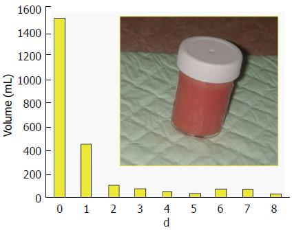
Figure 5 Insertion of a chest tube in the left hemithorax revealed 1500 mL of milky blood fluid (sample container).
The fluid gradually decreased in volume and became serous on day 7. The chest tube was removed on day 9.
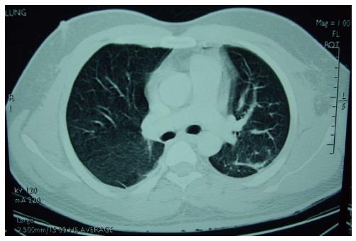
Figure 6 Computed tomography chest with intravenous contrast on day 10 showed complete resolution of the chylothorax.
- Citation: Idris K, Sebastian M, Hefny AF, Khan NH, Abu-Zidan FM. Blunt traumatic tension chylothorax: Case report and mini-review of the literature. World J Clin Cases 2016; 4(11): 380-384
- URL: https://www.wjgnet.com/2307-8960/full/v4/i11/380.htm
- DOI: https://dx.doi.org/10.12998/wjcc.v4.i11.380


