Copyright
©The Author(s) 2016.
World J Clin Cases. Nov 16, 2016; 4(11): 375-379
Published online Nov 16, 2016. doi: 10.12998/wjcc.v4.i11.375
Published online Nov 16, 2016. doi: 10.12998/wjcc.v4.i11.375
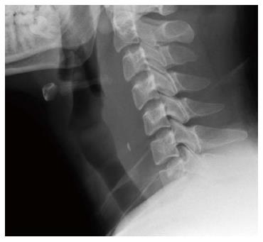
Figure 1 Radiopaque foreign body at the level of C5 vertebral body.
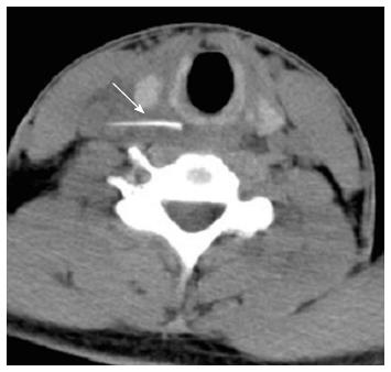
Figure 2 A thin sharp-ended foreign body embedded within the soft tissue of the right neck just below the vocal cord region (level C5/C6) which was close to the right common carotid artery (arrow).
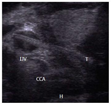
Figure 3 A linear foreign body seen coursing from the right thyroid lobe and run obliquely piercing the right common carotid artery with the tip of foreign body is abutting the internal jugular vein.
Small haematoma (H) seen between the right lobe and right common carotid artery. T: Thyroid lobe; CCA: Common carotid artery; IJV: Internal jugular vein.
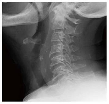
Figure 4 Radiopaque foreign body in a vertical position at C5-C6 level of vertebral body.
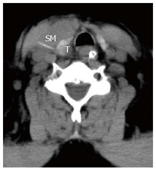
Figure 5 Linear dense foreign body was seen adjacent to the right thyroid lobe within the strap muscles.
The tip is seen within the inferior pole of right thyroid lobe (T). There was an intramuscular collection with air pockets seen in the adjacent strap muscles extending to submandibular area. SM: Strap muscles.
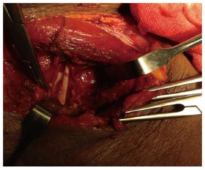
Figure 6 A 4 cm fish bone with a sharp jagged side located at the edge of the right thyroid lobe piercing through the medial aspect of the right sternocleidomastoid muscle.
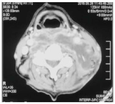
Figure 7 Fish bone at the level C3, measures 2.
5 cm in length (arrow) with hypodense collection at the retropharyngeal area extend from C1-C5 level, and left parapharyngeal area extending laterally and posteriorly to the left carotid space.
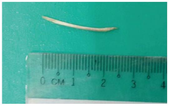
Figure 8 Fish bone measure 2.
5 cm embedded in the slough tissue medial to the posterior end of the sternocleidomastoid muscle.
- Citation: Johari HH, Khaw BL, Yusof Z, Mohamad I. Migrating fish bone piercing the common carotid artery, thyroid gland and causing deep neck abscess. World J Clin Cases 2016; 4(11): 375-379
- URL: https://www.wjgnet.com/2307-8960/full/v4/i11/375.htm
- DOI: https://dx.doi.org/10.12998/wjcc.v4.i11.375


