Copyright
©The Author(s) 2015.
World J Clin Cases. Jul 16, 2015; 3(7): 661-670
Published online Jul 16, 2015. doi: 10.12998/wjcc.v3.i7.661
Published online Jul 16, 2015. doi: 10.12998/wjcc.v3.i7.661
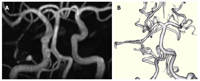
Figure 1 Three-dimensional time-of-flight magnetic resonance angiography.
A: Three-dimensional time-of-flight magnetic resonance angiography (3D-TOF-MRA) four years prior to admission; B: Volume rendering 3D-TOF-MRA study 1 year prior to admission, demonstrating two unruptured prenidal feeder artery aneurysms, one 8 mm × 3 mm fusiform proximal anterior inferior cerebral artery (AICA) aneurysm near the takeoff of the basilar artery and one 4.5 mm × 3 mm elliptical distal pedicle AICA aneurysm located at the entrance to the main inflow area of a small inconspicuous vascular nidus.
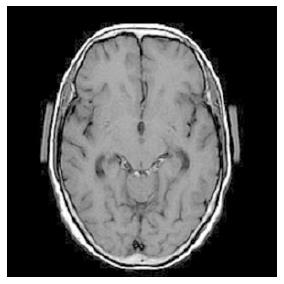
Figure 2 On admission, this 70-year-old woman presented with a severe headache, nausea and vomiting of several days duration.
T1-weighted magnetic resonance imaging demonstrated high intensity signals only in the quadrigeminal cistern, suggestive of early subacute subarachnoid hemorrhage.
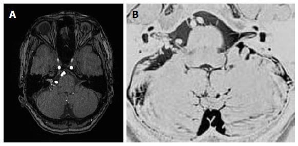
Figure 3 Magnetic resonance angiographic source imaging (A) and magnetic resonance cisternography (B) reveal a small (11.
4 mm × 4.2 mm) relatively compact nidus located in the cerebellopontine angle at the cisternal portion of the cranial nerves VII and VIII with extension into the internal auditory canal.
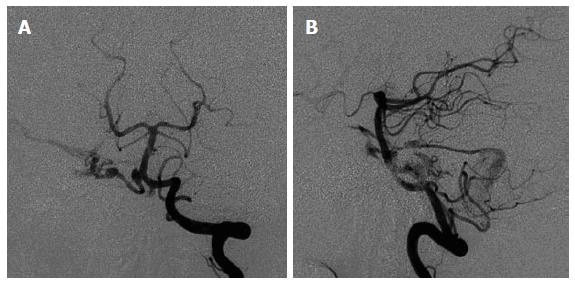
Figure 4 Anterioposterior (A) and lateral (B) views of cerebral angiogram for this patient showing the right anterior inferior cerebral artery as the predominant feeder supplying a small nidus with drainage into the right petrosal vein, superior petrosal sinus, and cavernous sinus [Spetzler-Martin grade III (S1V1E1)].
Figure 5 Intraoperative microscopic view.
A: Intraoperative microscopic view showing a right retrosigmoid craniotomy in the a right lateral oblique position which exposed a small nidus buried within the right cranial nerve VIII at the meatus of the internal auditory canal; B: View showing distal right anterior inferior cerebral artery feeding aneurysm, nidus-VIII cranial nerve complex, and draining petrous vein; C: Definitive clip application proximal to the aneurysm and partial electrocoagulation of the nidus and the inflow area just proximal to the nidus was performed.
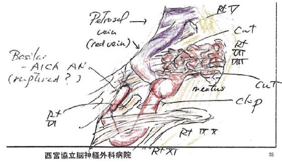
Figure 6 Intraoperative schematic illustration depicting anatomical structures and definitive clipping between the proximal and distal feeding anterior inferior cerebral artery aneurysms and partial electrocoagulation of both the nidus and the main inflow area to the nidus.
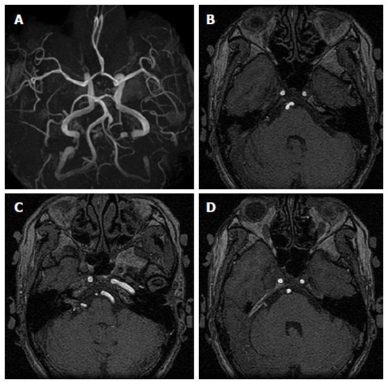
Figure 7 Preoperative three-dimensional time-of-flight magnetic resonance angiography showing proximal and distal right anterior inferior cerebral artery aneurysms (A), and magnetic resonance angiographic source imaging showing proximal right anterior inferior cerebral artery aneurysm (B), distal right anterior inferior cerebral artery aneurysm with relatively strong internal auditory canal nidus signal intensity (C), and right superior petrosal sinus venous shunt (D).
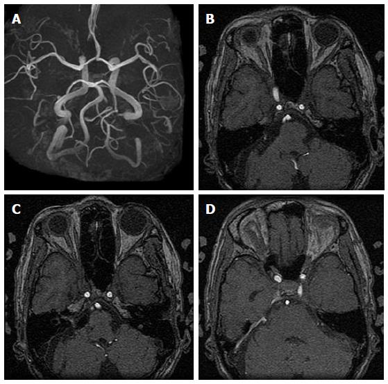
Figure 8 Postoperative three-dimensional time-of-flight magnetic resonance angiography (3D-TOF-MRA) and magnetic resonance angiographic source imaging showing anterior inferior cerebral artery (AICA) stump with disappearance of proximal and distal right AICA aneurysms (A, B), decreased intensity of right AICA and seemingly disappearance of proximal aneurysm, clip artifact at the entrance to the internal auditory canal and decreased intensity of intracanicular nidus (C), and persistence of prominent right superior petrosal sinus venous shunt (D).
- Citation: Tucker A, Tsuji M, Yamada Y, Hanabusa K, Ukita T, Miyake H, Ohmura T. Arteriovenous malformation of the vestibulocochlear nerve. World J Clin Cases 2015; 3(7): 661-670
- URL: https://www.wjgnet.com/2307-8960/full/v3/i7/661.htm
- DOI: https://dx.doi.org/10.12998/wjcc.v3.i7.661


