Copyright
©The Author(s) 2015.
World J Clin Cases. Jul 16, 2015; 3(7): 607-613
Published online Jul 16, 2015. doi: 10.12998/wjcc.v3.i7.607
Published online Jul 16, 2015. doi: 10.12998/wjcc.v3.i7.607
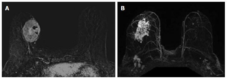
Figure 1 Magnetic resonance imaging phenotypes solid unifocal mass (A) and more diffuse non-mass enhancement (B).
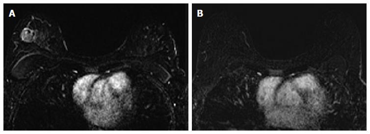
Figure 2 Forty-two years old woman with triple negative right breast cancer.
A: Baseline axial T1-weighted post-gadolinium fat-saturated magnetic resonance image demonstrates a 3.6 cm unifocal mass in the upper outer quadrant; B: Post-neoadjuvant chemotherapy magnetic resonance imaging demonstrates complete resolution of the mass seen previously. Surgical pathology demonstrates biopsy site changes and expected changes related to chemotherapy with no evidence of residual cancer.
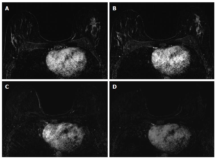
Figure 3 Thirty-seven years old woman with HR+ left breast cancer.
A and B: Baseline axial T1-weighted post-gadolinium fat-saturated magnetic resonance image demonstrates a speculated mass and contiguous non-mass enhancement extending posteriorly for a total of 7 cm of disease in the upper outer breast; C and D: Post-neoadjuvant chemotherapy magnetic resonance imaging demonstrates decrease in size and degree of enhancement of prior findings. Surgical pathology demonstrates 6.9 cm of invasive ductal carcinoma.
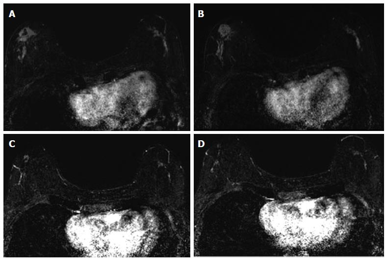
Figure 4 Sixty-four years old woman with bilateral HR+ breast cancer.
A and B: Baseline axial T1-weighted post-gadolinium fat-saturated magnetic resonance image demonstrates 3.2 cm irregular mass and contiguous non-mass enhancement (NME), spanning up to 7.2 cm, in the right central outer breast and 3.5 cm of clumped linear NME in the central outer left breast; C and D: Post-neoadjuvant chemotherapy magnetic resonance imaging demonstrates decrease in size of the right breast mass and NME. NME in the left breast demonstrates only mild improvement. Surgical pathology demonstrates 4.3 cm of residual disease on the right and 3.7 cm of disease on the left.
- Citation: Price ER, Wong J, Mukhtar R, Hylton N, Esserman LJ. How to use magnetic resonance imaging following neoadjuvant chemotherapy in locally advanced breast cancer. World J Clin Cases 2015; 3(7): 607-613
- URL: https://www.wjgnet.com/2307-8960/full/v3/i7/607.htm
- DOI: https://dx.doi.org/10.12998/wjcc.v3.i7.607


