Copyright
©The Author(s) 2015.
World J Clin Cases. Jun 16, 2015; 3(6): 525-532
Published online Jun 16, 2015. doi: 10.12998/wjcc.v3.i6.525
Published online Jun 16, 2015. doi: 10.12998/wjcc.v3.i6.525
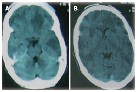
Figure 1 Cranial computed tomography brain showing axial views (A, B) with bilateral multifocal large hypodense lesions in the frontal, parietal, temporal and occipital lobes.
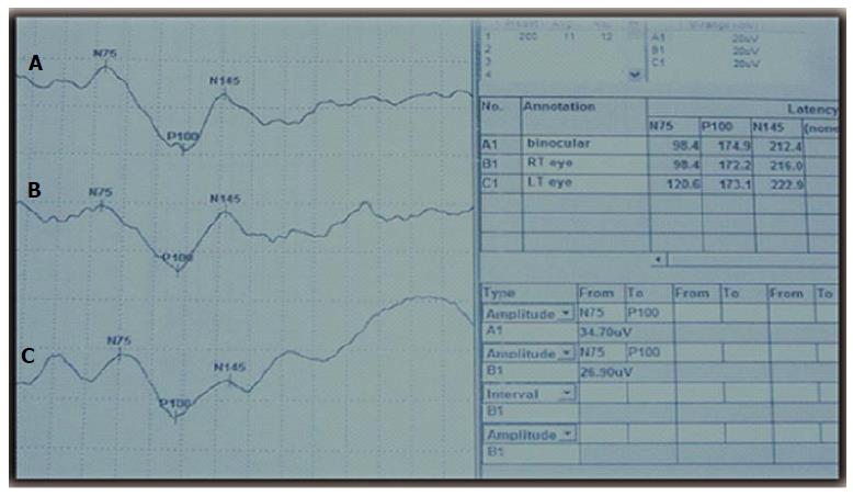
Figure 2 Visual evoked potentials showing prolonged absolute latencies of the P100 component of the right eye (A), left eye (B) and binocular (C).
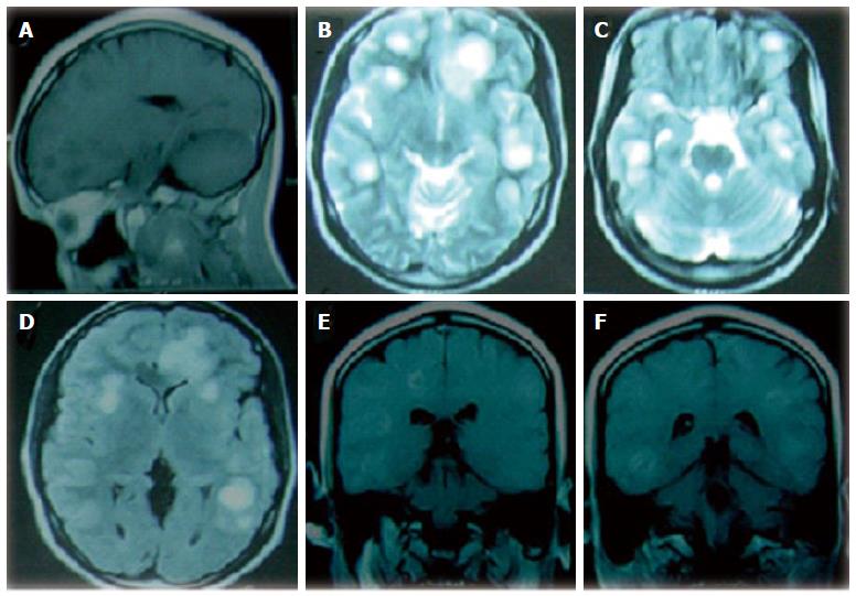
Figure 3 Cranial magnetic resonance imaging brain (on admission) showing (A) sagittal T1-weighted view with multiple hypointense lesions in the frontal and parietal regions, (B, C) axial T2-weighted images showing multifocal large hyperintense lesions in the frontal, parietal, temporal and occipital lobes, (D) axial fluid-attenuated inversion recovery-weighted image showing hyperintense lesions with minimal perifocal hypointense rim (edema), (E, F) coronal T1-weighted contrast enhanced views with patchy enhancement of the hypodense lesions in the temporal, parietal and frontal lobes bilaterally.
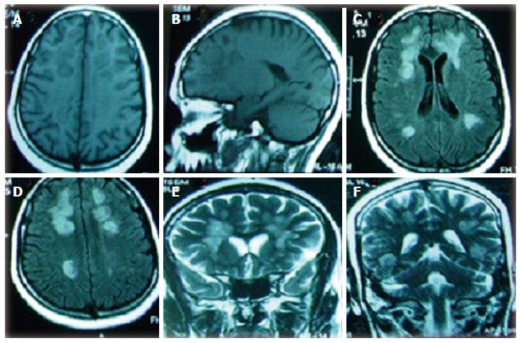
Figure 4 Cranial magnetic resonance imaging brain (after 6 mo of follow up) showing (A, B) axial and sagittal T1-weighted views with multiple hypointense lesions in the frontal, parietal, temporal and occipital regions, (C, D) axial fluid-attenuated inversion recovery-weighted images showing multifocal hyperintense lesions, (E, F) coronal T2-weighted images showing multifocal bilateral hyperintense lesions.
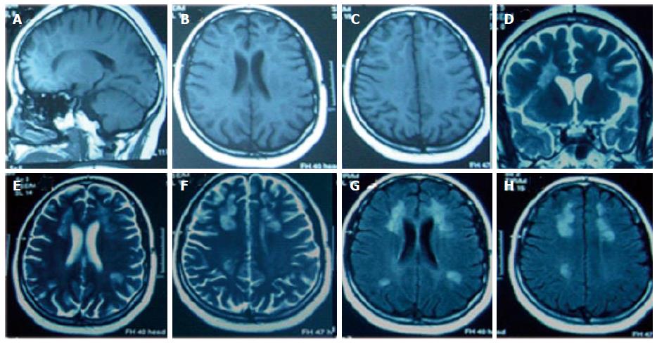
Figure 5 Cranial magnetic resonance imaging brain (after one year of follow up) showing (A, B, C) sagittal and axial T1-weighted views with bilateral multiple hypointense lesions in the frontal, parietal, temporal and occipital regions, (D, E, F) coronal and axial T2-weighted images showing multifocal bilateral hyperintense lesions, (G, H) axial fluid-attenuated inversion recovery-weighted images showing multifocal hyperintense lesions.
In all views, lesions had reduced sizes compared to those at 6 mo follow up.
- Citation: Hamed SA. Variant of multiple sclerosis with dementia and tumefactive demyelinating brain lesions. World J Clin Cases 2015; 3(6): 525-532
- URL: https://www.wjgnet.com/2307-8960/full/v3/i6/525.htm
- DOI: https://dx.doi.org/10.12998/wjcc.v3.i6.525


