Copyright
©The Author(s) 2015.
World J Clin Cases. May 16, 2015; 3(5): 457-461
Published online May 16, 2015. doi: 10.12998/wjcc.v3.i5.457
Published online May 16, 2015. doi: 10.12998/wjcc.v3.i5.457
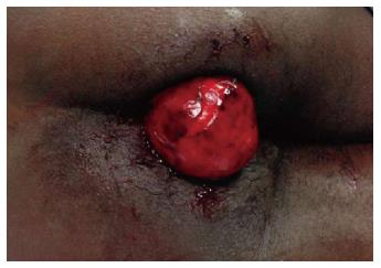
Figure 1 Protruding mass from anal canal in the emergency room (left lateral decubitus position).
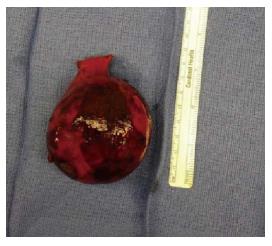
Figure 2 Gross view of the resected specimen.
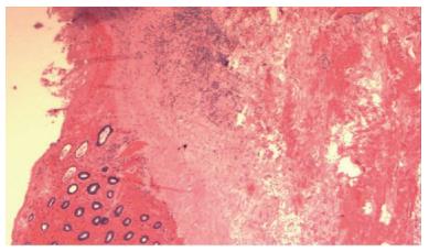
Figure 3 Histopathology (HE, 2 ×) showed colonic lipoma; the colonic mucosa (left side) has atrophy and ischemic necrosis with hemorrhage.
- Citation: Ghanem OM, Slater J, Singh P, Heitmiller RF, DiRocco JD. Pedunculated colonic lipoma prolapsing through the anus. World J Clin Cases 2015; 3(5): 457-461
- URL: https://www.wjgnet.com/2307-8960/full/v3/i5/457.htm
- DOI: https://dx.doi.org/10.12998/wjcc.v3.i5.457


