Copyright
©The Author(s) 2015.
World J Clin Cases. Apr 16, 2015; 3(4): 377-380
Published online Apr 16, 2015. doi: 10.12998/wjcc.v3.i4.377
Published online Apr 16, 2015. doi: 10.12998/wjcc.v3.i4.377
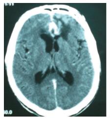
Figure 1 Computerized tomography scan of the year 2008.
Second episode of the intracerebral hemorrhage.
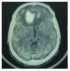
Figure 2 Computerized tomography scan of the year 1990.
First episode of intracerebral frontal hemorrhage (white arrow).
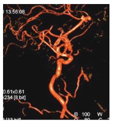
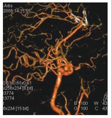
Figure 3 Preoperative digitally subtracted angiography.
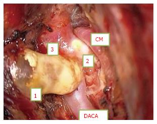
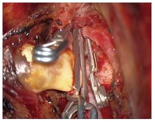
Figure 4 Intraoperative pre-clipping photo.
Numbers 1, 2 and 3 refer to respective lobes of the trilobed aneurysm. CM: Calloso-marginal; DACA: Distal anterior cerebral artery/pericallosal artery.
Figure 5 Intraoperative post-clipping photo.
Three separate clips exclude the three lobes of the aneurysm.
Figure 6 Postoperative digitally subtracted angiography showing preserved blood flow in the pericallosal artery and the exclusion of the aneurysm.
- Citation: Seferi A, Alimehmeti R, Rroji A, Petrela M. Saccular trilobed aneurysm of azygos anterior cerebral artery. World J Clin Cases 2015; 3(4): 377-380
- URL: https://www.wjgnet.com/2307-8960/full/v3/i4/377.htm
- DOI: https://dx.doi.org/10.12998/wjcc.v3.i4.377


